Abstraktbuch zum - Neurochirurgie der Universität
Werbung
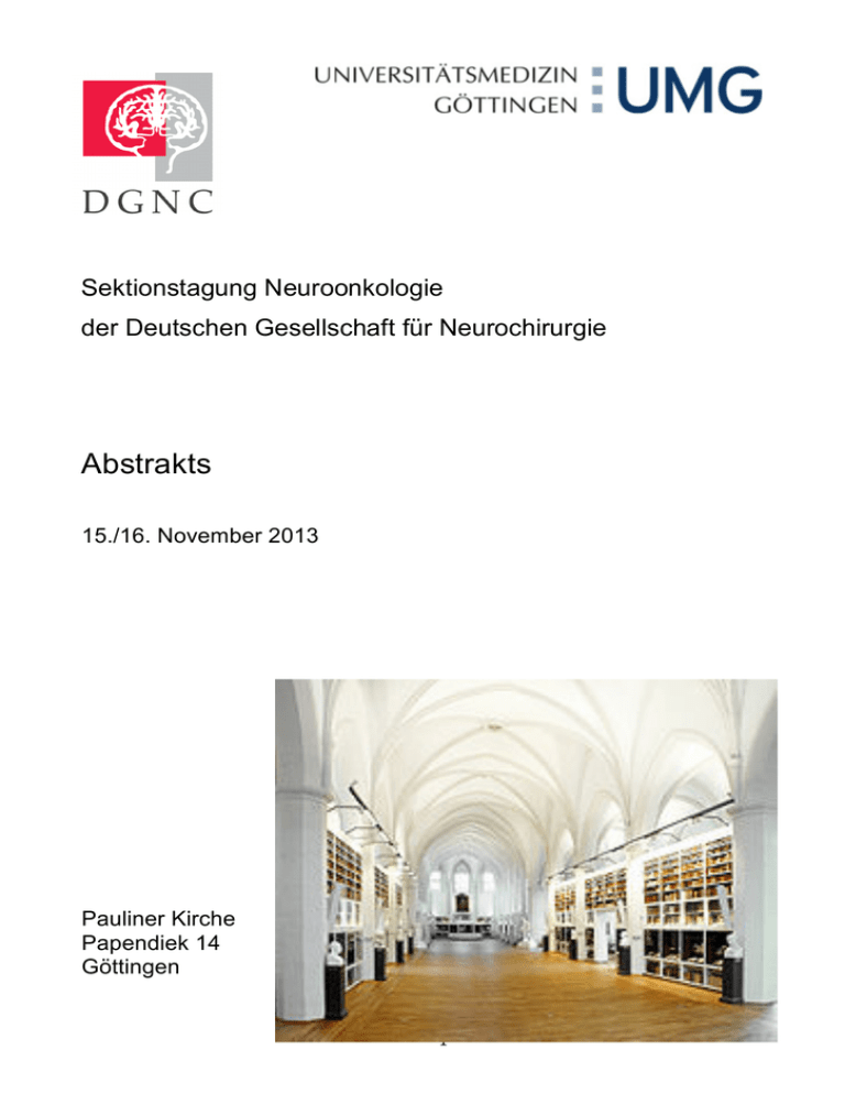
Sektionstagung Neuroonkologie der Deutschen Gesellschaft für Neurochirurgie Abstrakts 15./16. November 2013 Pauliner Kirche Papendiek 14 Göttingen 1 Administratives Wahl des besten Vortrages Wie in den letzten Jahren wird der beste Vortrag der Arbeitstagung der Sektion Neuroonkologie zur Jahrestagung der DGNC vom 11.-14. Mai 2014 nach Dresden eingeladen. Um eine transparente, unmittelbare Wahl des besten Vortrages zu gewährleisten werden wird diese über Doodle machen. Alle Teilnehmer müssen zu Registrierung eine Email mit angeben. Über diese Emailadresse wird ein Link zur Doodle Wahl verschickt. Die Wahl kann dann am besten über ein mobiles internettaugliches Gerät (Smartphone, Tablet, Notebook) erfolgen. Eine WLAN Einwahl wird mind. zur Wahl in der Paulinerkirche bereitstehen. Ferner werden wir Rechner zur Verfügung stellen, über die ebenfalls gewählt werden kann. Über den Link kann man sich wiederholt einwählen und bis zum Abschluss der Wahl, seine Entscheidung ändern. Es können pro Wahl mehrere Vorträge gewählt werden. Gewinner ist der Vortrag mit den meisten Nennungen. Das Ergebnis der Wahl wird unmittelbar zum Ende der Veranstaltung live durch Doodle generiert werden. Zertifizierung Die Ärztekammer Niedersachsen hat die Veranstaltung mit 10 Punkten als Fortbildungsveranstaltung zertifiziert. Bitte bringen sie ihre Bar-Code Aufkleber mit. Registrierung und Organisation PD Dr. Florian Stockhammer, [email protected] www.neurochirurgie-uni-goettingen.de Unterstützt durch 2 Programm Freitag 15.11.2013 12:00 Registrierung, Kaffee 13:00 Begrüßung Rohde (Göttingen) Simon (Bonn) Stockhammer (Göttingen) 13:15 - 14:27 Grenzen der Gliomresektion Vorsitz: Ernestus (Würzburg) 13:15 - 13:25 Gross total but not incomplete resection of GBM prolongs survival in the era of radiochemotherapy Friedrich-Wilhelm Kreth, N. Thon, M. Simon, M. Westphal, G. Schackert, G. Nikkhah, B. Hentschel, G. Reifenberger, T. Pietsch, M. Weller, J.-C. Tonn (München, Deutsches Gliomnetzwerk) 13:27 - 13:37 Besitzt die Einschätzung des Resektionsausmasses durch den Operateur prognostischen Charakter? Ist der Operateur besser als sein Ruf? Timo Behm, V. Rohde, F. Stockhammer (Göttingen) 13:39 - 13:49 Intraoperativer Ultraschall bei der Resektion vorbestrahlter Gliome Kay Mursch, F. Kalaji, W. Brück, M. Scholz, J. Behnke-Mursch (Bad Berka, Göttingen, Duisburg) 13:51 - 14:01 Influence of Surgery on Local Recurrence Pattern by GBM: Perceptions of a Single Centre Study Maria A.S. Zella, J. Baernreuther, M. Rapp, H.-J. Steiger, M. Sabel (Düsseldorf) 14:03 - 14:13 Brain metastasis: Infiltration of metastatic cells into the benign brain tissue is cancer-type dependent and correlates with poor prognosis Rene-B. Moringlane, A. Bleckmann, A. Mohr, H.A. Wolff, V. Rohde, C. Stadelmann, T. Pukrop, L. Siam (Göttingen) 14:15 - 14:25 Supramarginal resection of cerebral metastases: 1-year follow up Marcel A. Kamp, H. Sadat, M. Dibué, J. Kuzibaev, H.-J. Steiger, A. Szelényi, M. Rapp, M. Sabel (Düsseldorf) 14:30 - 15:00 Kaffeepause 3 15:00 - 16:24 Molekulargenetik Vorsitz: Simon (Bonn) 15:00 - 15:10 DNA methylation alterations in WHO grade II- and malignant pleomorphic xanthoastrocytoma Ramón Martínez, F. J. Carmona, V. Rohde, M. Kirsch, G. Schackert, W. Paulus, M. Esteller (Göttingen, Barcelona, Dresden, Münster) 15:12 - 15:22 The prognostic impact of IDH1 R132H mutation in malignant glioma treated by combined radiochemotherapy Antonia Horowski, T. Behm, Ch. Stadelmann, A. Barrentes, F. Behling, V. Rohde, Ch. Hartmann, F. Stockhammer (Göttingen, Hannover) 15:24 - 15:34 MACC1 reguliert die Migration und Invasion von Glioblastomzellen Carsten Hagemann, S. Fuchs, A. F. Kessler, P. Herrmann, J. Smith, T. Hohmann, U. Grabiec, Th. Linsenmann, F. Dehghani, R.-I. Ernestus, M. Löhr, U.Stein (Würzburg) 15:36 - 15:46 FTY720 (fingolimod) has strong antiproliferative effects in glioblastoma cells in vitro Malgorzata A.Kolodziej, B. Al-Barim, M. Stein, M. Reinges, E. Uhl, K. Quint (Giessen) 15:48 - 15:58 Das mitotische Kontrollpunktprotein MPS1 als neues Ziel bei der Therapie des Glioblastoms Almuth F. Kessler, T. Linsenmann, R.-I. Ernestus, M. Löhr, T. Würdinger, C. Hagemann (Würzburg, Amsterdam) 16:00 - 16:10 Auswirkung der Notch-Signalweg Aktivierung beim primären Glioblastom Nicolai El Hindy, Y. Zhu, Ph. Dammann, U. Sure (Essen) 16:12 - 16:22 Expression von Karyopherin a2 and Chromosome Region Maintenance Protein 1 bei Meningeompatienten: neue Biomarkers für Tumorrezidiv und Malignisierung. Konstantinos Gousias , P. Niehusmann, M. Simon (Bonn) 16:30 - 17:00 Kaffeepause 4 17:00 - 18:12 Bildgebung von Gliomen Vorsitz: Stockhammer (Göttingen) 17:00 - 17:10 Dynamic 18FET-PET in suspected WHO grade II gliomas defines distinct biological subgroups with different clinical courses Niklas Thon, M. Kunz, L. Armbruster, N. L. Jansen, S. Eigenbrod, S. Kreth, J. Lutz, R. Egensperger, A. Giese, J. Herms, M. Weller, H. Kretzschmar, J.-C. Tonn, C. la Fougère, F. Kreth (München, Zürich) 17:12 - 17:22 Dynamic 18FET-PET in newly diagnosed astrocytic low grade glioma identifies high risk patients Bogdana Suchorska, N. L. Jansen, V. Wenter, S. Eigenbrod, C. Schmid-Tannwald, A. Zwergal, M. Niyazi, M. Drexler, P. Bartenstein, O. Schnell, J.C. Tonn, N. Thon, C. la Fougère, F. W. Kreth (München) 17:24 - 17:34 Prognostic value of 18FET-PET on the clinical course in newly diagnosed glioblastoma Bogdana Suchorska, N. L. Jansen, J. Linn, H. Kretzschmar, H. Janssen, S. Eigenbrod, M. Simon, G. Pöpperl, F. W. Kreth, C. la Fougere, M. Weller, and J.C. Tonn (München, Deutsches Gliomnetzwerk) 17:36 - 17:46 ALA-derived fluorescence in patients suffering from radionecrosis after glioblastoma treatment Hosai Sadat, M. Dibue, J. Kuzibaev, H.-J. Steiger, M. Rapp, M. A. Kamp, M. Sabel (Düsseldorf) 17:48 - 17:58 Multiphoton microscopy: a novel tool for label-free imaging of brain tumors Ortrud Uckermann, R. Galli, K. Geiger, G. Steiner, G. Schackert, M. Kirsch (Dresden) 18:00 – 18:10 Intraoperative speech mapping by cortical thermography and multivariate data analysis Yordan Radev, N.Hoffmann, J.Hollmach, C.Schnabel, S.Sobottka, E.Koch, G.Schackert, G.Steiner, M.Kirsch (Dresden) 18:12 - 18:22 Sinusvenenthrombosen nach Entfernung intrakranieller Tumoren. Eine seltene aber ernste Komplikation Florian Geßler, M. Bruder, S. Dützmann, J. Quick, V. Seifert, C. Senft (Frankfurt/M) 19:30 gemeinsames Abendessen 5 Samstag 16.11.2013 08:45 Kaffee 09:00 -11:00 freie Themen I Vorsitz: Krex (Dresden) 09:00 - 09:10 Does 5-ALA have a Radiosensitizing Effect? Andrei Nemes, V. Senner, B. Greve, W. Stummer, Ch. Ewelt (Münster) 09:12 - 09.22 Sind Radiochirurgie (RS) und stereotaktische Radiotherapie (SRT) bei hirneigenen Tumoren und speziell beim Rez.-Glioblastom sinnvoll? Klaus Hamm, H.-U. Herold, G. Surber, R. Gerlach, S. Rosahl (Erfurt) 09:24 - 09:34 Survival improvement in patients with glioblastoma multiforme-treated in a specialized neurooncology center in comparison to an oncology outpatient center Claudia Schlimper, R. Lefering, A. Brunn, F. Weber (Köln) 09:36 - 09:46 Beneficial impact of a neuro-oncological setting in outpatient care for patients with newly diagnosed glioblastoma Naureen Keric, B. Tomoschat, T. Behm, M. Renovanz, Ch. Richter, V. Rohde, A. Giese, H. Ch. Bock (Mainz, Göttingen) 09:48 - 09:58 Glioma patient management and follow-up pattern of care organized by an all-in-one database application Naureen Keric, M. Renovanz, A. Giese, H. Ch. Bock (Mainz, Göttingen) 10:00 - 10:10 Erfassung der Lebensqualität von Patienten mit primären und rezidivierenden Glioblastomen: eine monozentrische Studie Julia Steinmann, M. A. S. Zella, M. Rapp, H.-J. Steiger, M. Sabel (Düsseldorf) 10:12 - 10:22 Neurokognitive Testung bei Gliompatienten Carolin Weiß, L. Seiler, A. K. Rehme, R. Goldbrunner (Köln) 10:24 - 10:34 Projektvorstellung: Psychoonkologisches Screening neurochirurgischer Patienten- Einfluss der Diagnose, des Screeningszeitpunktes sowie Validierung eines geeigneten Screeningstools Marion Rapp, K. Hoffmann, M. Wallocha, H.J. Steiger, M. Sabel (Düsseldorf) 10:36 - 10:46 Hyperprolactinemia in young females with questionable lesion in sellar imaging. Indication for surgery? Christian Ewelt, M. Richters, A. Nemes, B. Fischer, W. Stummer (Münster, Bochum) 10:50 - 11:00 Administratives 11:00 - 12:00 Mittagspause, Imbiss 6 12:00 - 13:20 freie Themen II Vorsitz: Weyerbrock (Freiburg) 12:00 - 12:10 Cerebrospinal fluid shunting in glioblastoma Bujung Hong, M. Abdallat, H. Heissler, Ph. Ertl, M. Nakamura, J. K. Krauss (Hannover) 12:12 - 12:22 Klinische Verbesserung durch ventrikuloperitonealen Shunt bei Patienten mit kommunizierendem Hydrozephalus und malignen Gliomen. Christin Clajus, T. Behm, V. Rohde, F. Stockhammer (Göttingen) 12:24 - 12:34 TMS-basierte Cortexkartierung und Faserbahndarstellung als präklinischer Standard in der Gliomchirurgie ? Carolin Weiß, I. Tursunova, V. Neuschmelting, A. Eisenbeis, G. Stoffels, A. OrosPeusquens, Ch. Nettekoven, Ch. Grefkes, R. Goldbrunner (Köln) 12:36 - 12:46 Salivary melatonin concentration and sleep patterns before and after pinealectomy in patients with pineocytoma WHO°I Melatoninkonzentration im Speichel und Schlafmuster vor und nach Pinealektomie bei Patienten mit Pineozytom WHO°I H. Slawik; Michael Behr, M. Stoffel, B. Meyer, J. Lehmberg; M. Wiegand, S. M. Krieg (München) 12:48 - 12:58 Verbessert der Schnellschnitt die Diagnostische Sicherheit von Stereotaktischen Biopsien? Martin Misch, G.-H. Schneider, A. Koch, F. Prall, K. Lange, F. Stockhammer (Berlin, Rostock, Göttingen) 13:00 - 13:10 Biopsy of cortical cerebral lesions. Comparison of navigated free-hand versus frame-based stereotactic biopsy Awad Aleid, M. Misch, V. Rohde, F. Stockhammer (Göttingen, Berlin) 13:15 Wahl des besten Vortrages 13:25 Schlußworte 13:30 Ende der Veranstaltung 7 Gross total but not incomplete resection of GBM prolongs survival in the era of radiochemotherapy Friedrich-Wilhelm Kreth1*, N. Thon1*, M. Simon2, M. Westphal3, G. Schackert4, G. Nikkhah5, B. Hentschel6, G. Reifenberger7, T. Pietsch8, M. Weller9, J.-C. Tonn1 1 Department of Neurosurgery, University of Munich LMU, Campus Großhadern, Munich, Germany; 2Department of Neurosurgery, University of Bonn Medical Center, Bonn, Germany; 3 Department of Neurological Surgery, University Medical Center Hamburg-Eppendorf, Hamburg, Germany; 4Department of Neurosurgery, Technical University of Dresden, Dresden, Germany; 5Department of Stereotactic Neurosurgery, University of Freiburg, Freiburg, Germany; 6Institute for Medical Informatics, Statistics and Epidemiology, University of Leipzig, Leipzig, Germany; 7Department of Neuropathology, Medical Faculty, Heinrich-Heine University Düsseldorf, Düsseldorf, Germany; 8Institute of Neuropathology, University of Bonn Medical Center, Bonn, Germany; 9Department of Neurology, University Hospital Zurich, Zurich, Switzerland. Background This prospective multicenter study assessed the prognostic influence of extent of resection as compared to biopsy only in a contemporary patient population with newly diagnosed glioblastoma. Patients and methods Histology, O6-methylguanine-DNA methyltransferase (MGMT) promoter methylation status and clinical data were centrally analysed. Survival analyses were performed with the Kaplan-Meier method. Prognostic factors were assessed with proportional hazards models. Results Of 345 patients, 273 underwent open tumor resection and 72 biopsies; 125 patients had gross total resections (GTR) and 148 patients incomplete resections. Surgery-related morbidity was lower after biopsy (1.4% vs. 12.1%, p=0.007). 64.3% of patients received radiotherapy (RT) plus chemotherapy (CT), 20.0 % RT alone, 4.3% CT alone, and 11.3% best supportive care as initial treatment. Patients ≤ 60years and with a Karnofsky performance score (KPS) ≥ 90 were more likely to receive RT plus CT (p<0.01). Median OS (PFS) ranged from 33.2 months (15.0 months) for patients with MGMT methylated tumors after GTR and RT plus CT to 3.0 months (2.4 months) for biopsied patients receiving supportive care only. Favorable prognostic factors in multivariate analysis for OS were age ≤ 60 years (HR=0.52; p<0.001), preoperative KPS ≥ 80 (HR=0.55; p<0.001), GTR (HR=0.60; p=0.003), MGMT promoter methylation (HR=0.44; p<0.001), and RT plus CT (HR=0.18, p<0.001); patients undergoing incomplete resection did not better than those receiving biopsy only (HR=0.85; p=0.31). Conclusions The value of incomplete resection remains questionable. If GTR cannot be safely achieved, biopsy only might be used as an alternative surgical strategy. 8 Besitzt die Einschätzung des Resektionsausmasses durch den Operateur prognostischen Charakter? Ist der Operateur besser als sein Ruf? Timo Behm, V. Rohde, F. Stockhammer Klinik für Neurochirurgie, Universitätsmedizin Göttingen Objekt Das Ausmaß der primären Resektion in der Behandlung des Glioblastoma multiforme stellt einen gewichtigen prognostischen Faktor dar. Standard für die Beurteilung ist heute die früh- postoperative cMRT. Bei retrospektiven Betrachtungen sind in vielen Kollektiven diese MRT- Untersuchungen nicht vorhanden und die Angabe des Resektionsausmasses beruht lediglich auf der Einschätzung des Operateurs. In dieser Untersuchung soll gezeigt werden, in welchem Ausmaß diese Einschätzung auch in Bezug auf andere prognostische Faktoren (Alter, KPS, adjuvante Therape) verlässlich und konklusiv erscheint. Methode Einschlusskriterien in dieser retrospektiven Untersuchung sind: histologisch gesichertes Glioblastom behandelt in der Universitätsmedizin Göttingen im Zeitraum von 1998 bis 2007, Alter ≥ 18 Jahre, postoperativ erfolgte Radiatio (± adjuvante Chemotherapie) oder konkomitante Radiochemotherapie, kein früh-postoperatives cMRT, keine intraoperative 5-ALAFluoreszenz. Die Einschätzung des Resektionsausmasse erfolgte in die Gruppen vollständige Resektion (GTR), subtotale Resektion (STR) und Biopsie. Ergebnis 249 Patienten wurden eingeschlossen, das mediane Alter beträgt 61,8 Jahre (20-85 J.), hiervon sind 99 Patienten ≥ 65 J., 150 Patienten < 65 J., 83 Patienten mit einem KPS < 70%, 166 Patienten mit einem KPS ≥ 70%, 103 Patienten erhielten postoperativ eine konkomitante Radiochemotherapie, 146 erhielten postoperativ eine Radiatio ± adjuvante Chemotherapie. Das mediane Überleben beträgt bei Biopsie 8,2 Monate bei STR 11,3 Monate und bei GTR 13 Monate (p < 0,0038). Auch die Subgruppenanalyse (Alter < 65 J. vs. ≥ 65 J., konkomitante Radiochemotherapie, KPS ≥ 70%) zeigt signifikante Vorteile für die Resektion. Lediglich in den Subgruppen KPS < 70% und Radiatio ± Chemotherapie kann kein signifikanter Vorteil nachgewiesen werden. Zusammenfassung Die intraoperative Einschätzung des Resektionsausmaßes durch den Operateur erscheint in der retrospektiven Analyse prognostisch. 9 Intraoperativer Ultraschall bei der Resektion vorbestrahlter Gliome Kay Mursch1, F. Kalaji1, W. Brück2, M. Scholz3, J. Behnke-Mursch 1 Neurochirurgische Klinik, Zentralklinik, Bad Berka, 2Institut für Neuropathologie, Universitätsmedizin, Georg August Universität, Göttingen, 3Neurochirurgische Klinik, Klinikum Wedau, Duisburg Fragestellung Intraoperativer Ultraschall (IOUS) ist ein nützliches Instrument zur Navigation und Resektionskontrolle bei neurochirurgischen Operationen. Bekanntermassen ist aber die Abgrenzung von Tumoren zum umgebenden bereits bestrahlten Gewebe schwierig. Wir untersuchten, 1. ob IOUS die Navigation zu bestrahlten Tumor ermöglicht 2. ob IOUS die Resektionsränder definieren kann. Patienten und Methoden Siebzehn bereits an einem Gliom operierte und bestrahlte Patienten wurden untersucht. Wärend der Operation eines Tumorrezidivs wurde IOUS verwendet und die Bilder videodokumentiert. Wir entnahmen zwei Proben: 1. aus für den Chirurgen makroskopisch unzweifelhaftem Tumorgewebe 2. Gewebe aus dem Resektionsrand, für den Chirurgen unsichere Zuordnung. Resultate Eine postoperative semiquantitative Analyse der Ultraschallbilder zum Zeitpunkt der Probeentnahme ergab, 1. dass alle 17 erste Proben aus makroskopisch sicherem Tumorgewebe histologisch auch Tumorgewebe entsprachen und vom Ultraschallaspekt her hyperechogen und als pathologisch abgrenzbar waren. 2. dass im Resektionsrand eine Unterscheidung zwischen Tumorgewebe/Infiltrationszone/tumorfreiem vorbestrahltem Gewebe durch IOUS nicht möglich ist. Sowohl in isoechogenem Gewebe als auch in hyperechogenem Gewebe fanden sich alle o.g. Histologien (Tabelle). Ultraschallbild Isoechogen Leicht hyperechogen Histologie Tumor (n=) 1 5 Infiltration (n=) 2 4 Kein Tumor (n=) 1 4 Zusammenfassung IOUS führt auch in bestrahltem Gewebe zum Tumor. Eine Resektionskontrolle nach dem Ultraschallaspekt ist in vorbestrahltem Gewebe nicht sicher möglich. 10 Influence of Surgery on Local Recurrence Pattern by GBM: Perceptions of a Single Centre Study Maria A.S. Zella, J. Baernreuther, M. Rapp, H.-J. Steiger, M. Sabel (Düsseldorf) Objective With the introduction of the fluorescence-guided resection technique, the rate of complete resections of the contrast enhancing part of the Glioblastoma multiforme (GBM) significantly increased. In spite of that, due to infiltrating growth pattern, the typical GBM recurrences after open resection occur locally, usually within 2 cm from the border of the original lesion. In the present work the authors are interested to know whether a more aggressive local surgical treatment might influenced the rate of local recurrences. Patients and methods Inclusion criteria for this single centre, retrospective study were: Complete resection of GBM at first diagnosis (complete resection was defined as no residual contrast enhancement (CE) in early postoperative MRI T1 sequences plus CE [<72h]), followed by at least the concomitant phase of Stupp protocol. For all patients included in the study recurrence was histologically confirmed. At recurrences, patients were divided into two groups according to cerebral MRI imaging (T1 sequences plus CE): patients with a local recurrence (within 2 cm from the primary tumor's margins), patients with a distant recurrence (≥ 2 cm from the primary tumor margins). Furthermore possible pattern of recurrence were analyzed and found to be either per continuitatem, subependymal (in clear contact with ependyma), along fiber tracts, along CSF dissemination, de novo or multifocal. In addition, survival data were analyzed. Results: 53 patients who underwent complete tumor resection for a primary GBM between January 2007 and December 2011, followed by at least the concomitant phase of Stupp protocol were identified. At first recurrence, 48 patients (86%) presented a local recurrence, 8 patients (14%) a distant recurrence (2 patients (25%) with CSF disseminations, 2 patients (25%) with de novo manifestation, 3 patients (38%) with recurrence along fibre tracts and 1 patient (12%) with subependymal relapse were identified). In 3 cases more than one recurrence pattern was identified. At second recurrence, 10 patients (71%) presented a local recurrence, 4 patients (29%) a distant recurrence (1 patient (25%) with CSF disseminations, 2 patients (50%) with de novo manifestation, and 1 patient (25%) with subependymal relapse). First recurrence: median survival for patients with local recurrence (48 cases) was 28.9 months (range 15.2-42.7) versus 16.3 months (range 9.1 – 23.6) for the patients with distant recurrence (8 cases) without significant difference between the groups (P-value: 0.18 Log Rank, 0.21 Tarone-Ware).Second recurrence: median survival for patients with local recurrence (10 cases) was 21.0 months (range 15.1 - 26.8) versus 43.0 months (range 5.5-80.4) for patients with distant recurrence (4 cases) without significant difference between the groups (P-value: 0.13 Log Rank, 0.17 TaroneWare). Conclusions: Despite aggressive surgical treatment with no residual CE, the vast majority of recurrences still occur locally (86% of local recurrence by 1st Recurrence, 71% by 2. Recurrence). No significant differences in OAS or PFS between Local and Distant Recurrence Groups were notable. Even though its limits (restrospective non randomized study and lack of genetical infos), this work demonstrates that if functionality is not compromised, even more aggressive “supramarginal” treatment might be considered to improve tumor local control. 11 Brain metastasis: Infiltration of metastatic cells into the benign brain tissue is cancer-type dependent and correlates with poor prognosis Rene-B. Moringlane, A. Bleckmann, A. Mohr, H.A. Wolff, V. Rohde, C. Stadelmann, T. Pukrop, L. Siam (Göttingen) Objective Brain metastases of various cancers have unfavorable prognosis. However, the treatment haven ́t changed in recent decades; and individualized therapy is far from the current modalities. In contrast, neither the biological differences of the primary tumors are really considered nor the resection status of solitary cerebral metastases needs to be determined, like in liver metastasis. The latter is one consequence of the ongoing debate whether metastatic cells infiltrate the adjacent brain tissue. So far the only prospective biopsy study demonstrated no evidence of infiltrating metastatic cells beyond the resection margin while two autopsy studies indicate the opposite. Prospective study design To clarify this contradictory data up to ten biopsies per patient of the resection cavity wall were taken directly after completed gross total resection of brain metastasis and analyzed by suitable IHC for infiltrating tumor cells afterwards. Results Twenty-six patients were included in this prospective study and 127 biopsies were taken. In 42 biopsies (33.1%) infiltrated tumor cells were detectable, 16 of the 26 patients (61.5%) revealed infiltrated tumor cells at least in one biopsy sample. Furthermore, the infiltration depends on the primary tumor histology and has significant impact on prognosis. Conclusion First, our results prove the existence of infiltrating metastatic cells. Moreover, the infiltration has prognostic value and is tumor type dependent. At this time where even the standard therapies of brain metastasis are highly scrutinized our results are very useful in decision-making, in particular dependent on the tumor type, and for the design of more biological tailored study concepts. 12 Supramarginal resection of cerebral metastases: 1-year follow up Marcel A. Kamp, H. Sadat, M. Dibué, J. Kuzibaev, H.-J. Steiger, A. Szelényi, M. Rapp, M. Sabel Neurochirurgische Klinik, Heinrich-Heine-Universität, Düsseldorf, Germany Background Although being standard therapy, microsurgical circumferential stripping of intracerebral metastases is often insufficient in preventing local tumor control. Supramarginal resection may improve local tumor control. Therefore, we retrospectively analyzed a series of patients with cerebral metastases in eloquent and non-eloquent areas for neurological outcome and tumor control during 1-year follow-up. Methods A retrospective analysis was performed for all patients, who underwent supramarginal resection as awake surgery with intraoperative cortical and subcortical stimulation, MEPs and SSEPs, for a cerebral metastasis between 03/2009 and 09/2012. Pre- and postsurgical neurological status was assessed by the NIH Stroke Scale. Permanent deficits were defined by persistence after 3 months observation time. Results Supramarginal resection of cerebral metastases in eloquent and non-eloquent brain areas were performed in 30 patients with a mean age of 58 years (33 – 83 y). 4/30 patients (13.3%) had a new transient postoperative neurological deficit, which improved within a few days due to a supplementary motor area (SMA) syndrome (median preoperative NIHSS: 2, 0 – 12, standard deviation, SD: 2.5; median postoperative NIHSS: 2, 0 – 9, SD: 2.2). 4/30 pts. (13.3%) developed local recurrences and 8 patients (26.6%) distant recurrences. For these patients, mean progression-free survival was 12 months (2 – 45 months). Conclusion Supramarginal resection of cerebral metastases with intraoperative monitoring is a feasible tool for metastases in eloquent areas. Despite aggressive resection we observed no permanent postoperative neurological deficits. Furthermore, supramarginal resection might achieve a better tumor control as frequency of local recurrences is lower as in recent published series. Further studies are needed in order to analyze the benefit of this method in achieving better tumor control. 13 DNA methylation alterations in WHO grade II- and malignant pleomorphic xanthoastrocytoma Ramón Martínez, F. J. Carmona, V. Rohde, M. Kirsch, G. Schackert, W. Paulus, M. Esteller (Göttingen, Barcelona, Dresden, Münster) Pleomorphic xanthoastrocytoma (PXA) is a rare WHO grade II benign tumor accounting for less than 1% of all astrocytomas. Malignant transformation into PXA with anaplastic features, corresponding to WHO grade III, is unusual and correlates with poorer outcome of the patients. Using a DNA methylation platform, we have quantified the DNA methylation level of 1,505 CpG dinucleotides (807 genes) of WHO grade II and III PXA and compared to normal brain tissue and glioblastoma multiforme samples. Increasing DNA promoter hypermethylation events were observed to be parallel to malignant transformation of PXA. The dissection of DNA methylation alterations in malignant PXA uncovered common traits also affecting GBM. Specifically, we have found a set of epigenetically disrupted genes (FZD9, CD81, TES, HOXA5, ASCL2, DIO3, HCK and MOS) potentially associated with PXA malignant progression. Further studies assessing DNA methylation changes in this setting might unveil epigenetic mechanisms underlying PXA pathogenesis and provide biomarkers useful for clinical management, especially in benign forms at high risk to become malignant. 14 The prognostic impact of IDH1 R132H mutation in malignant glioma treated by combined radiochemotherapy Antonia Horowski1, T. Behm1, Ch. Stadelmann2, A. Barrentes2, F. Behling2, V. Rohde1, Ch. Hartmann3, F. Stockhammer1 (Göttingen, Hannover) 1 Klinik für Neurochirurgie, Universitätsmedizin Göttingen, 2Institut für Neuropathologie Universitätsmedizin Göttingen, 3Institut für Neuropathologie, Medizinische Hochschule Hannover Objective In astrocytic malignant glioma mutation of isocitrate dehydrogenase (IDH) 1 R132H has been reported to have more impact on survival than histological diagnosis. However, anaplastic gliomas in that latter study had either radiotherapy or chemotherapy, but no combined treatment like the glioblastomas compared to. In our institution anaplastic astrocytomas haven been treated likewise glioblastomas. We set up a retrospecive analysis investigating the role of IDH1 mutation in anaplastic astrocytomas and glioblastomas equally treated. Methods All patients with anaplastic astrocytoma or glioblastoma who received combined radiochemotherapy as first-line treatment and immunohistochemical staining for IDH1 R132H antibody were included. Results 187 patients were elegible for this analysis. 130 patients (70%) deceased. Histology revealed 27anaplastic astrocytoma and 160 glioblastomas. IDH1 R132H mutation was present in 16 patients, haboring 9 anaplastic astrocytoma and 7 glioblastoma. Median survival in glioblastoma patients was 14.0 and in anaplastic astocytomas 52.0 months (p<.0001). Median survival in glioblastoma with IDH1 R132H mutation sustained unfavorable compared to anaplastic astrocytomas with IDH wildtype (15.0 vs. 38.1 months, p=0.033, log rank). Conclusion Histology has superior impact on survival compared to IHD1 R132H mutation, when patients are equally treated. These results indicate that patients with IDH1 wildtype anaplastic astrocytoma might profit from combined treatment. 15 MACC1 reguliert die Migration und Invasion von Glioblastomzellen Carsten Hagemann1, S. Fuchs1, A. F. Kessler1, P. Herrmann2, J. Smith2, T. Hohmann3, U. Grabiec3, Th. Linsenmann1, F. Dehghani3, R.-I. Ernestus, M. Löhr1, U. Stein2 1 University of Würzburg, Department of Neurosurgery, Tumorbiology Laboratory, JosefSchneider-Str. 11, 97080 Würzburg, 2Experimental and Clinical Research Center, Charité University Medicine Berlin, at the Max-Delbrück-Center for Molecular Medicine, Robert-RössleStr. 10, 13125 Berlin, 3Martin-Luther-University Halle-Wittenberg, Department of Anatomy and Cell Biology, Grosse Steinstr. 52, 06108 Halle (Saale) Hintergrund Metastasis-associated in colon cancer-1 (MACC1) ist ein unabhängiger prognostischer Indikator für die Metastasenbildung und das metastasenfreie Überleben beim Kolonkarzinom. Obwohl Glioblastome (GBM) nur sehr selten außerhalb des Zentralnervensystems (ZNS) Metastasen bilden, ist ihr Invasions- und Migrationsverhalten jenem von metastatischen Zellen anderer Tumorentitäten sehr ähnlich. Vor kurzem zeigten wir eine Assoziation der mRNA- und Proteinkonzentrationen von MACC1 mit der WHO-Gradierung von Gliomen. Die MACC1 Expression erlaubte hierbei sowohl eine Diskriminierung zwischen nicht progredienten und rezidivierenden Astrozytomen WHO Grad 2 als auch zwischen primären und sekundären GBM. Des Weiteren förderte eine transiente Überexpression von MACC1 die Proliferation und Migration von GBM-Zellen. Methoden Es wurde ein Datamining öffentlicher Datenbanken durchgeführt, die Microarrayund The Cancer Genome Atlas (TCGA) Daten zugänglich machen, um die Überexpression von MACC1 zu bestätigen. Die Migration primärer Zellkulturen von GBM-Patienten wurde mit Hilfe von Sphäroid-Migrationsassays untersucht. Außerdem wurden die Migration und die Invasion MACC1 transfizierter U138 Zellen unter Einfluss des Met-Inhibitors Crizotinib in real-time (xCELLigence) gemessen. Die Fähigkeit dieser Zellen, Tumoren zu bilden, wurde schließlich in organotypischen Hippokampus-Schnittkulturen (organotypic hippocampal slice cultures; OHSC) von Mäusen bestimmt. Ergebnisse Die MACC1 Überexpression konnte basierend auf den großen Probenzahlen der Datenbanken bestätigt werden und korrelierte mit der Amplifikation von Chromosom 7. Die endogene Expression von MACC1 variierte allerdings in aus GBM-Tumoren gewonnenen Primärzellen. Dabei korrelierten hohe Expressionslevel positiv mit der Zellmigration in einem Sphäroidassay. In OHSC förderte MACC1 die Migration, Invasion und Tumorbildung von GBMZellen, während der Met-Inhibitor Crizotinib eine Reversion auf den Basallevel bewirkte. Die Überexpression von MACC1 verursachte eine Steigerung der Met-Expression. Dies könnte ein erster Hinweis sein, dass auch in GBM MACC1 Effekte über die Regulation der Met-Expression vermittelt werden. Schlussfolgerung MACC1 beeinflusst die Migration, Invasion und Tumorbildungsfähigkeit von GBM-Zellen möglicherweise über eine Regulation des Hepatozytenwachstumsfaktor(hepatocyte growth factor; HGF) Rezeptors Met. Eine Inhibition von MACC1 könnte einen neuen therapeutischen Ansatz für die Hemmung der GBM-Zellmigration und -Invasion darstellen. 16 FTY720 (fingolimod) has strong antiproliferative effects in glioblastoma cells in vitro Malgorzata A. Kolodziej, B. Al-Barim, M. Stein, M. Reinges, E. Uhl, K. Quint (Giessen) Objective The sphingosine-1-phosphate (S1P) pathway is involved in cell survival, growth, migration and angiogenesis and is overexpressed in glioma. It’s effects are mediated through G-protein coupled receptors signaling to the PI3K/Akt, Rho/ROCK and Ras/ERK/MAPK cascades. We and others have previously shown that sphingosine kinase 1 and S1P-receptor 1 expression influence survival of glioma patients. FTY720 (fingolimod) is an S1P analogue with promising effects in glioblastoma. Here we investigate the antiproliferative effects of FTY720 versus temozolomide (TMZ) in glioblastoma cell lines and investigate the modulation of downstream signaling pathways. Methods Glioblastoma cell lines A172, G28 and U87 were incubated with 5, 10, 25 and 50 µM FTY720 or temozolomide (TMZ) for 24 to 72 hours and proliferation measured using an MTT assay and the xCELLigence real-time cell analyzer system, an impedance based cell proliferation and viability system. IC50 values for FTY720 were calculated at 72 hours and compared to TMZ effects. Gene expression of downstream pathways was quantified by realtime quantitative PCR. Results Already after 24 hours, 10 µM FTY720 reduced viable A172 cells to less than 5% and after 72 hours, 5 µM FTY720 to 22.6% and 10 µM to 2.3% of untreated controls. Similar dosedependent results were obtained for G28 and U87 cells with viable cells below 2% at 72 hours using 50 µM FTY720. IC50 at 72 hours of FTY720 incubation was 4.6 µM in A172 cells, 17.3 µM in G28 and 25.2 µM in U87 cells, respectively. Its effects surpassed those of TMZ, which did not reduce viable cell counts below 50% in any cell line even at 50 µM. In qPCR, FTY720 lead to decrease of downstream pathway components Akt1, MAPK1, PRKCE, RAC1 and ROCK1 to values of 0.2 in G28 and U87 but to increase of those components to value of 1.2 in A172, respectively at FTY720 for 72 hours. Conclusion FTY720 has strong antiproliferative effects on glioblastoma cells at micromolar concentrations and greatly surpasses TMZ effects. FTY720 suppresses Akt1, MAPK1, PRKCE, RAC1 and ROCK1 on the mRNA level and has antiproliferative effects in low concentrations. Therefore, it might be a promising targeted drug in this cancer. 17 Das mitotische Kontrollpunktprotein MPS1 als neues Ziel bei der Therapie des Glioblastoms Almuth F. Kessler1, T. Linsenmann1, R.-I. Ernestus1, M. Löhr1, T. Würdinger2, C. Hagemann1 1 University of Würzburg, Department of Neurosurgery, Tumorbiology Laboratory, JosefSchneider-Str. 11, 97080 Würzburg, 2 VU University Medical Center, De Boelelaan 1117, 1081 HV Amsterdam, The Netherlands Hintergrund Die chemotherapeutische Behandlung des Gliobastoma multiforme WHO°IV (GBM) zielt unter anderem auf eine DNS-Schädigung während der Mitose, welche zur „mitotic catastrophe“ und zum Zelltod führen soll. Eine solche Substanz mit antimitotischem Wirkmechanismus ist Vincristin, dessen Anwendung in der neuroonkologischen Therapie fest etabliert ist. Der mitotische Kontrollpunkt überprüft die korrekte Anheftung der Spindelfasern an die Chromatiden, erlaubt eine Reparatur der chemotherapeutisch gesetzten Schäden und stellt dadurch die korrekte Chromosomentrennung sicher. Ein Schlüsselprotein dieses Kontrollpunktes ist die Kinase Monopolar Spindle 1 (MPS1). Eine Beeinflussung des mitotischen Kontrollpunktes könnte zu einem besseren Ansprechen der Chemotherapie bei GBM-Patienten führen. Methoden Die mRNA-Expression verschiedener Proteine des mitotischen Kontrollpunktes, einschließlich MPS1, wurde mittels semiquantitativer RT-PCR in GBM-Zelllinien und Tumorproben von GBM-Patienten analysiert. Immunhistochemische Färbungen wurden zum Nachweis der MPS1-Proteinexpression in 24 Astrozytomen WHO°II (LGA) und 14 GBM (10 primäre und 4 sekundäre) durchgeführt. Der WHO-Grad des Tumors und die Überlebenszeit der Patienten wurden mit den MPS1-Expressionsdaten aus öffentlich zugänglichen GliomDatenbanken korreliert. Der Effekt einer MPS1-Inhibition wurde in Kombination mit Vincristingabe in einem orthotopen GBM-Mausmodell analysiert (n = 3 bis 7 Mäuse/Gruppe). Ergebnisse MPS1 war im GBM-Tumorgewebe deutlich überexprimiert. Dies korrelierte positiv mit dem Tumorgrad und negativ mit der Überlebenszeit der Patienten (P < 0.001). Gliompatienten mit hoher MPS1-Expression (n = 203) hatten eine mediane Überlebenszeit von 487 Tagen, während Patienten mit mittlerer MPS1-Expression (n = 140) im Median 858 Tage überlebten. Die 2-Jahresüberlebensrate dieser Gruppen lag bei 35 % bzw. 56 %. Im orthotopen GBM-Mausmodell führte die MPS1-Hemmung in Verbindung mit Vincristinbehandlung zu einer Tumorvolumenreduktion und verlängerten Überlebenszeit. Schlußfolgerung Die MPS1-Inhibition sensibilisiert GBM-Zellen gegenüber antimitotischen Chemotherapeutika. Dies legt MPS1 als neuen therapeutischen Angriffspunkt in der GBMTherapie nahe. Die synergistische MPS1-Hemmung könnte zum Beispiel eine Dosisreduktion des neurotoxischen Vincristins erlauben. Der Stellenwert von MPS1 als prognostischer Marker sollte in prospektiven Studien weiter evaluiert werden. 18 Auswirkung der Notch-Signalweg Aktivierung beim primären Glioblastom Nicolai El Hindy, Y. Zhu, Ph. Dammann, U. Sure Klinik für Neurochirurgie, Universitätsklinikum Essen Hintergrund Glioblastome sind aggressiv wachsende Tumore, welche sich durch Angiogenese, Nekrose und Therapieresistenz auszeichnen. Ein deregulierter Notch- Signalweg wurde mit der Formation und Progression verschiedener Malignome in Zusammenhang gebracht. Die vorgelegte Studie untersucht die Aktivierung wesentlicher Komponenten des Notch-Signalwegs im primären GBM und seine Assoziation mit vaskulären und klinischen Parametern. Methodik Die Hauptkomponenten des Notch-Signalwegs wurden mittels Real-Time PCR, Western-Blot und immunhistochemischen Untersuchungen im GBM- (n=26) und Kontroll-HirnGewebe (n=11, nach epilepsie-chirurgischen Eingriffen) untersucht. Eine Analyse der Gefäßarchitektur, sowie der mittleren Gefäßdichte fand nach Färbung von Mikrogefäßen mittels Laminin statt. An klinischen Parametern floss der MGMT-Promotor Methylierungs-Status sowie die peritumorale Ödemformation ein. Ergebnisse Die mRNA Level von Dll4, Jagged1, Notch1, Notch4, Hey1, Hey2, Hes1 und VEGF waren im GBM im Vergleich zum Kontrollgewebe um das 3.12-, 3.58-, 3.37-, 5.77-, 4.89-, 3.13-, 6.62-, und 32.57-fache erhöht. Die Western-Blot Untersuchung zeigte eine 4-, 3.7-, und 45.6fache Überexpression von Dll4, Notch1 und Hey1. Dies ging mit einer Unterexpression von PTEN und einer Überexpression von p-AKT und VEGF einher. Die immunhistochemischen Untersuchungen lokalisierten Dll4 und Notch1 auf Endothelzellen, Mikroglia/Makrophagen, Tumorzellen und Astrozyten. Verantwortlich für die Überexpression war allerdings nur eine Subgruppe der untersuchten GBM, in der die Überexpression der Notch-Signalweg Komponenten signifikant mit einer geringeren Gefäßdichte und einer produktiveren Gefäßarchitektur, vom klinischen Aspekt mit einer verstärktem Ödembildung und der positiven MGMT-Promotor Methylierung assoziiert war. Schlußfolgerung Die Überexpression der Notch-Signalweg Komponenten wurde in einer Untergruppe primärer GBM gefunden und war mit vaskulären und klinischen Parametern assoziiert. Dies weist auf einen potentiellen therapeutischen Nutzen dieses Signalwegs bei Patienten mit GBM und Aktivierung des Notch-Signalwegs hin. 19 Expression von Karyopherin a2 and Chromosome Region Maintenance Protein 1 bei Meningeompatienten: neue Biomarkers für Tumorrezidiv und Malignisierung. Konstantinos Gousias1 , P. Niehusmann2, M. Simon1 Abteilung für Neurochirurgie 1 und Abteilung für Neuropathologie 2, Universitätsklinik Bonn, Sigmund-Freud-Strasse 25, 53105 Ziel Die Karyopherin-Protein-Familie besteht aus den Importinen und Exportinen und spielt eine wichtige Rolle beim nukleozytoplasmatischen Transport. Erhöhte Werte von Karyopherin a2 (KPNA2) (Importin) und Chromosome Region Maintenance Protein 1 (CRM1) (Exportin) korrelieren mit einer schlechten Prognose bei infiltrativen Gliomen sowie bei verschiedenen soliden Tumoren wie Mamma-, Ovarial-, Uterus-, Nieren- und Prostatakarzinomen sowie Melanomen und Osteosarkomen. Das Ziel dieser Studie war, die o.g Karyopherine als neue Biomarker auch für Meningeome WHO-Grad I-III zu evaluieren. Material und Methoden Wir analysierten semiquantitativ mittels Immunohistochemie (Labeling index) die nukleäre Expression von KPNA2, CRM1 und MIB1 in 108 Primär- (44 WHO-Grad I, 47 WHO-Grad II, 17 WHO-Grad III) und 13 Rezidivmeningeome. Zur statistischen Analyse der Ergebnisse wurden Standardverfahren eingesetzt. Ergebnisse Die Expression von KPNA2 (p<0,001) und CRM1 (p=0,002) korrelierte signifikant mit dem histologischen Grad (p<0,001). Weiterhin, korrelierte die Expression von KPNA2 mit der proliferativen Aktivität (MIB1-Index, p<0,001). Rezidivtumore exprimierten im Vergleich zu Primärtumoren deutlich mehr KPNA2 (p=0,045). Die multivariate Analyse für die gesamte Kohorte sowie für die atypischen Meningeome identifizierte eine niedrige KPNA2- und CRM1Expression (definiert als immunhistochemischer Labeling index <5% für KPNA2 und < 60% für CRM1, jeweils Median) als unabhängige Prognosefaktoren für das progressionsfreie Überleben. Schlussfolgerungen Zusammenfassend zeigen die Ergebnisse, dass die Expression von KPNA2 und CRM1 sich potentiell als neue diagnostische und prognostische Biomarkers auch für Meningeome eignen. 20 Dynamic 18FET-PET in suspected WHO grade II gliomas defines distinct biological subgroups with different clinical courses Niklas Thon1*, M. Kunz1*, L. Armbruster1, N. L. Jansen2, S. Eigenbrod3, S. Kreth4, J. Lutz5, R. Egensperger5, A. Giese3, J. Herms3, M. Weller6, H. Kretzschmar3, J.-C. Tonn1, C. la Fougère2, F. Kreth1 1 Department of Neurosurgery; 2Department of Nuclear Medicine, 3Center for Neuropathology and Prion Research; 4Department of Anaesthesiology, 5Department of Clinical Radiology, Ludwig-Maximilians-University, Munich, Germany; 6Department of Neurology, University of Zurich, Switzerland Purpose In suspected grade II gliomas, the existence of three distinct patterns of time-activitycurves (TAC) in O-(2-[18F]fluoroethyl)-1-tyrosine (18FET) positron emission tomography (PET) has recently been uncovered: homogeneous intra-tumoral increasing (decreasing) TAC with corresponding low-grade (high-grade) histology, and a combination of both increasing and focal decreasing TAC with corresponding low-grade and high-grade histology. This prospective study analyzed whether these patterns correlate with distinct biological tumor subtypes and different prognoses. Experimental Design 18FET-PET guided biopsies were used for stepwise histopathological evaluation. Molecular-genetic evaluation included O6-methylguanine-DNA methyltransferase (MGMT) promoter methylation, isocitrate dehydrogenase (IDH1/2) mutational and 1p/19q codeletion status. Progression-free survival (PFS) was estimated with the Kaplan-Meier method. Prognostic factors were obtained from multivariate regression models. Results 98 adult patients were included. Histology indicated 54 low-grade and 44 high-grade gliomas. Homogeneous increasing, focal decreasing, and homogeneous decreasing TAC were seen in 51, 19, and 28 patients, respectively. The corresponding one-year (two-years) PFS were 92% (85%), 89% (50%), and 50% (28%), respectively (p=0.0001). The areas with focal decreasing TAC covered 4-66% of the T2/FLAIR-hyperintense tumor volumes. IDH1/2 mutations were more frequent in tumors with homogeneous increasing (90%) and focal decreasing (74%) TAC, but were rare in those exhibiting homogeneous decreasing TAC (14%; p<0.001). Overall, TAC patterns, IDH1/2 mutational and 1p/19q codeletion status were powerful prognostic factors. Conclusions The compelling prognostic impact of metabolic profiles in suspected grade II gliomas offers perspectives for stratified diagnostic and therapeutic strategies. Highly targeted surgical interventions guided by metabolic imaging are recommended to avoid undergrading and undertreatment 21 Dynamic 18FET-PET in newly diagnosed astrocytic low grade glioma identifies high risk patients B. Suchorska1, N. L. Jansen2 , V. Wenter2, S. Eigenbrod3, C. Schmid-Tannwald4 A. Zwergal5, M. Niyazi6, M. Drexler2, P. Bartenstein2, O. Schnell1, J. C. Tonn1, N. Thon1, C. la Fougère2 and F. W. Kreth1 Departments of 1Neurosurgery, 2Nuclear Medicine, 3Neuropathology, 4Institute for Clinical Radiology, 5Department of Neurology, 6Department of Radiation Oncology, Ludwig-MaximiliansUniversity Munich Background The clinical course of gliomas WHO grade II (low grade gliomas, LGG) varies considerably depending on the histological subtype, biomarkers, patient’s age and tumor location. In order to identify further imaging factors which might be associated with outcome, we investigated the value of dynamic 18FET-PET in newly diagnosed LGG. Patients and Methods 83 patients with newly diagnosed LGG and dynamic FET-PET prior to histopathological assessment were retrospectively investigated. FET-PET analysis comprised a qualitative visual classification of lesions (FET-negative vs. FET-positive), the assessment of semi-quantitative parameters (maximal and mean tumor uptake, biological tumor volume) and a kinetic analysis (increasing vs. decreasing time-activity-curve (TAC)). PET parameters were correlated with progression-free and overall survival (PFS and OS) and with time to malignant transformation (TTM). Results Histopathological analyses revealed 59 astrocytomas WHO II and 24 oligodendroglial LGG WHO II. During follow-up (median time 37 months), 32/83 patients experienced tumor progression, of whom 20 presented with malignant transformation. Median PFS was 39.2 months, median TTP was not yet reached (mean 58.2 months). None of the semiquantitative parameters was associated with the clinical outcome; FET-negative LGG did not have a better prognosis than FET-positive gliomas. In contrast, patients with a decreasing TAC had a significantly shorter PFS and TTM in astrocytomas (p≤0.001), but not in oligodendrogliomas. Conclusions FET-negative LGG should not be considered as slowly growing gliomas with low metabolic activity. Decreasing TAC was shown to be associated with an unfavourable prognosis in astrocytomas WHO II. Thus, dynamic acquisition of 18FET-PET might help to identify more aggressive tumors in order to optimize a personalized treatment. 22 Prognostic value of 18FET-PET on the clinical course in newly diagnosed glioblastoma Bogdana Suchorska1*, N. L. Jansen2*, J. Linn3, H. Kretzschmar4, H. Janssen3, S. Eigenbrod4, M. Simon5, G. Pöpperl6, F. W. Kreth1, C. la Fougère2, M. Weller7, and J.C. Tonn1 (Deutsches Gliomnetzwerk) Departments of 1Neurosurgery, 2Nuclear Medicine, 3Neuroradiology and 4Neuropathology Ludwig-Maximilians-University Munich, 5Department of Neurosurgery, University of Bonn, 6 Department of Nuclear Medicine, Katharinenhospital Stuttgart and 7Department of Neurology, University Hospital Zurich, Switzerland Background Aim of this prospective longitudinal study conducted within the framework of the German Glioma network was to identify static and dynamic O-(2-[(18)F] fluoroethyl)-1-tyrosine PET (18FET-PET) derived imaging biomarkers in glioblastoma (GB) patients. Patients and Methods 92 patients with newly diagnosed GB eligible for radiochemotherapy (RCx) were included in this prospective study; diagnosis was obtained by biopsy (n=46) or resection (n=46). 18FET-PET and MRI scans were done prior to surgery, following RCx and after three cycles of temozolomide. At each timepoint, biological tumor volume (BTV), maximal 18 FET uptake as ratio to background (SUVmax/BG), tumor uptake kinetics (TAC) were obtained. Overall survival (OS) was the primary endpoint, the progression-free survival (PFS) was a secondary endpoint. ROC analyses were done to determine optimal cut-off values of 18FETPET parameters for survival outcome. To identify predictors for OS/PFS, Cox regression analysis was performed. Results Biological tumor volume (BTV) before RCx turned out to be the most important 18FETPET derived imaging biomarker, which was independent from MGMT promoter methylation and clinical prognostic factors: patients with smaller BTV did significantly better. BTV interacts with the mode of surgery: in the resection group, BTV gained prognostic relevance only after tumor surgery, whereas the BTV prior to any treatment was associated with prognosis in the biopsy group. 18FET time-activity-curves (TAC) before treatment and their changes after RCx were significantly related to outcome: patients with initially increasing TAC and those exhibiting a switch from decreasing to increasing TAC after RCx experienced longer progression-freesurvival. Conclusions BTV and TAC pattern represent important 18FET-PET-derived imaging biomarker in GB. Evaluation of TAC pattern after RCx allows early assessment of treatment response. The prognostic impact of BTV in the resection group depends on volume reduction by microsurgery 23 ALA-derived fluorescence in patients suffering from radionecrosis after glioblastoma treatment Hosai Sadat, M. Dibue, J. Kuzibaev, H.-J. Steiger, M. Rapp, M. A. Kamp, M. Sabel Department of Neurosurgery, Medical Faculty, Heinrich-Heine-University, Düsseldorf, Moorenstraße 5, D-40225 Düsseldorf, Germany. Background 5-Aminolevulinic acid (5-ALA) fluorescence guided resection of recurrent malignant glioma is a surgical standard procedure in many neuro-oncologic centers and was considered to be as reliable as for primary malignant gliomas. An important differential diagnosis of glioma recurrence is a pseudoprogression. Purpose of the present retrospective analysis was to analyse 5-ALA induced fluorescence (AIF) behaviour in tissue histopathologically classified as pseudoprogression after adjuvant treatment of a glioblastoma. Methods A retrospective analysis was performed for all patients suffering from malignant glioma who underwent surgical resection of pseudoprogression without recurrent tumor proven by frozen section. The presence of AIF in the tumor was intraoperatively estimated by the surgeon, using the categories no, faint and strong AIF. Results Between 2007 and 2013, a total of 17 patients underwent AIF-guided surgical resection of a pseudoprogression without recurrent tumor tissue. Mean age was 57.2 y (38 – 75y). Pretreatement was Stupp-Protocol in 15 patients. 2 patients received chemotherapy with temozolamid. Mean time between first glioblastoma resection and pseudoprogression surgery was 20.4 month (1-102m, SEM: 6.9). Totally, 2 patients (11.8%) showed no, 5 patients (29.4%) a strong and 10 patients faint AIF (58.8%). Conclusions Almost 30% patients of patients with histopathologically proven pseudoprogression without recurrent tumor after adjuvant treatment of a glioblastoma showed a strong AIF. Therefore, AIF should be carefully valued in the case of pseudoprogression. 24 Multiphoton microscopy: a novel tool for label-free imaging of brain tumors Ortrud Uckermann1, R. Galli2, K. Geiger3, G. Steiner2, G. Schackert1, M. Kirsch1 1 Klinik und Poliklinik für Neurochirurgie, Universitätsklinikum Carl Gustav Carus, Technische Universität Dresden, 2Klinisches Sensoring und Monitoring, Medizinische Fakultät, Technische Universität Dresden, 3Neuropathologie, Institut für Pathologie, Universitätsklinikum Carl Gustav Carus, Technische Universität Dresden Introduction In-vivo imaging of brain tumors is attracting increasing attention as a potential intra-operative tool for improving tumor border delineation. Coherent anti-Stokes Raman spectroscopy (CARS) is a non-invasive, label-free imaging technique that visualizes tissue morphology by exploiting chemical contrast. Through a non-linear interaction between ultrashort pulsed near-infrared laser beams and molecular vibrations, CARS addresses the biochemical composition of the tissue. It can be combined with other modalities that probe intrinsic tissue signals, e.g. endogenous two-photon excited fluorescence (TPEF) and second harmonic generation (SHG), to build a multimodal image. CARS tuned to the vibrations of C-H molecular groups visualizes the lipid distribution, while TPEF and SHG add information on cell morphology, vessels and extracellular matrix. Methods and Results The potential of the technique for histopathology and diagnosis was investigated by imaging fresh samples and cryosections of different types of human nervous tissue (n = 15) and experimental brain tumors in a mouse model (glioblastoma n = 8; metastasis of breast cancer n=4; melanoma metastasis n=4). For excitation of TPEF and SHG a ps laser (780 nm) was used; for induction of the CARS process a second ps laser source (1005 nm) was overlaid in time and space to resonantly excite C-H molecular vibrations in the sample. Multiphoton microscopy of cryosections provided exhaustive information of the tumor morphology. The tumor tissue could be discerned from the surrounding normal brain independent of the type of tumor (primary or secondary) and tumor´s properties like vascularization or proliferation. In the mouse model, the tumor border was defined with cellular resolution by reduction of CARS signal intensity to 61 % (glioblastoma), 71 % (melanoma) and 68 % (breast cancer), reflecting the lower lipid content within the tumor. Micrometastases infiltrating normal tissue (size 50 – 200 µm) were identified in glioblastoma and melanoma. Thanks to CARS imaging addressing lipids, we were able to identify grey and white matter and to locate fibre tracts. Additionally, axonal and cell morphology were retrieved without staining. Several tumors – Glioblastoma, neurinoma, pituitary adenoma – could be distinguished from the normal tissue on the basis of the alterations in morphology and composition. Necrotic areas were characterized by lipid droplets (imaged by CARS) fibrotic areas and transformed blood vessels network were detected thanks to SHG-acitive collagen. Conclusion All primary or secondary brain tumors investigated were characterized by a lower intensity of the CARS signal, therefore offering a simple tool for objective tumor detection. The combination of techniques allows imaging of native unstained tissue comparable to H&E staining. Multiphoton microscopy has the potential to become a technology routinely used intraoperatively for inspection and by neuropathology for assessment of brain tumors equivalent to frozen section. 25 Intraoperative speech mapping by cortical thermography and multivariate data analysis Yordan Radev1, N. Hoffmann2, J. Hollmach2, C. Schnabel2, S. Sobottka1, E. Koch2, G. Schackert1, G. Steiner2, M. Kirsch1 1 2 Klinik und Poliklinik für Neurochirurgie, Universitätsklinikum Carl Gustav Carus, Dresden, Klinisches Sensoring und Monitoring, Medizinische Fakultät, Technische Universität Dresden Introduction In awake craniotomies, identifying the areas involved in speech production is essential for a safe surgical procedure. Currently, mapping of the speech production region is performed by direct cortical stimulation during speech tasks. So far, no method of intraoperative visualization of cortical activity has been established. Thermography, which is based on measuring the infrared radiation of an object, is a safe, non-invasive, label-free technique with a long history of use in medicine. Neuronal activity is coupled to an increased tissue metabolism which is known to cause an increase in cerebral perfusion. Since the temperature of the exposed brain cortex correlates to perfusion, it may be possible to identify active areas by creating a cortical thermal map. In our study, we recorded cortical temperature during speech tasks and performed multivariate data analysis to evaluate the thermal activity patterns of the exposed brain surface. Materials and Methods In four awake craniotomies performed in patients bearing gliomas adjacent to the presumed Broca’s area, sequences of thermal images of the exposed cortex were recorded while the patients performed speech tasks. A highly-sensitive digital infrared camera was used to image cortical temperature distribution after craniotomy and exposure of the brain. The camera has a spectral range of 7,5 - 14 µm that corresponds to a thermal range of -40 to 1200°C, it reaches thermal resolution of 0,03K at 30°C and a spatial resolution of 150 µm per pixel at minimum distance. Preprocessing and multivariate data analysis was performed on thermal image data using the MATLAB package. The recorded thermographic images were matched to preoperative radiological images using Brainlab and Amira software applications as well as to the results of the direct cortical stimulation. Results In two of the four recorded thermographic sequences, we detected an activity pattern in the basal frontal lobe near the Sylvian fissure that mirrored the course of the speech tasks, i.e. an increase in temperature during speech and a decrease during rest. Using anatomical landmarks such as gyri, sulci and cortical vessels, the thermograms were matched to an intraoperative photograph and, consequently, to a 3D model of the patient’s brain. The areas that displayed the described thermal pattern corresponded to the basal motor cortex and Broca’s area and caused aphasia in the patient when directly stimulated. 26 Conclusions By using time-resolved intraoperative thermographic imaging of the brain cortex and multivariate analysis of the recorded data, we were able to detect functional activity in the basal motor cortex and Broca’s region during awake craniotomies. This is the first time to visualize the area activated in speech production, delineating Broca’s area next to an infiltrating tumor. Interestingly, the data did not show significant overlap to the pre-operative verbgeneration fMRI. By achieving a fast, reliable and artifact-free imaging of cortical activity, infrared thermography may become a useful tool for intraoperative visualization of functionally active cortical areas and thus increase the safety of neurosurgical procedures. 27 Sinusvenenthrombosen nach Entfernung intrakranieller Tumoren. Eine seltene aber ernste Komplikation Florian Geßler, M. Bruder, S. Dützmann, J. Quick, V. Seifert, C. Senft Klinik für Neurochirurgie, Johann Wolfgang Goethe-Universität Frankfurt am Main, Schleusenweg 2-16, 60528 Frankfurt am Main; [email protected] Objective Postoperative thromboembolic events occur frequently in patients harboring intracranial tumors. Especially removal of meningiomas or gliomas is known to be associated with hypercoagubility. The occurrence of sinus vein thrombosis (SVT) is a rare complication, and there is limited information on incidence, optimal treatment regimen and outcome. Methods From January 2004 to December 2012, a total of 8054 procedures were analyzed. Postoperative SVT was assessed radiologically. Patient and surgery related data (age, histology, tumor localization, patient positioning), as well as postsurgical clinical data, treatment and outcome were assessed. Results Postoperative SVT was diagnosed in 15 patients (0.2%; m:f 1:2). A single sinus thrombosis was present in 7 patients (46.7%), while 8 patients (53.3%) displayed SVT in multiple sinuses. Patients experiencing SVT suffered from meningioma in 7 cases (46.7%), glioblastoma in 1 case (6.7%), while 7 patients (46.6%) had other histologic diagnoses. Known risk factors for the onset of SVT or venous thrombo-embolism (VTE) were present in 4 patients (26.7%). A traumatic lesion of a sinus was reported in 10 patients (66.7%). Diagnosis was obtained in routine postoperative imaging in the vast majority of cases (n=12, 80%) rather than because of the development of new symptoms. Occurence of SVT was significantly more frequent in patients suffering from a posterior fossa lesion (p<0.001) and patients operated on in a semi-sitting position (p<0.001). Half-therapeutic anticoagulation was initiated directly in patients with diagnosis of SVT on p.o. day 1 or 2. If diagnosed later on, full heparinization was intitiated. Under anticoagulation, 2 patients had relevant hemorrhages and two patients developed ischemic strokes. After 3 months, a good outcome (mRS <2) was seen in 9 patients diagnosed with SVT. Two patients have died from complications within follow-up. Conclusion We provide information about the incidence, risk factors and outcome of postoperative SVT after craniotomy for intracranial tumors. While the incidence of postsurgical SVT is low, patient positioning and localization of pathologies seem to play a role in the occurrence of SVT. Good outcome may be achieved with cautious use of anticoagulation in SVT treatment. 28 Does 5-ALA have a Radiosensitizing Effect? Andrei Nemes1, V. Senner1, B. Greve2, W. Stummer3, Ch. Ewelt3 1 Institute of Neuropathology, Pottkamp 2 48149 Münster, 2 Department of Radiotherapy, AlbertSchweitzer-Str. 33 48149 Münster and 3 Department of Neurosurgery, Albert-SchweitzerCampus 1, Building A1 48149 Münster, University Hospital Munster Emitting red light and the generation of reactive oxygen species are two physical reactions of protoporphyrin IX (PPIX) upon irradiation with different wavelength of light. These two mechanisms are widely used either as an intraoperative tumor marker or to directly "attack" different kind of tumor entities. But what happens if you combine intracellular PPIX accumulation with another sort of photons, with X-rays or standard radiotherapy. Does 5aminolevulinic acid (5-ALA), despite its effect as a lightsensitizer, have a radiosensitizing effect? To address this hypothesis we investigated the combination of 5-ALA treatment and radiotherapy of different intensities in a human glioblastoma cell line and thereafter in glioblastoma slice cultures. The human glioblastoma cell line U373 was treated with and without 100 µg µL-1 5-ALA for 6 hours. Irradiation was performed with 6MeV photons of a linear accelerator (Varian Medical Systems, Palo Alto, CA, USA) and a dose rate of 4.8 Gy min-1. The cells were irradiated with 2.5, 4, 5 and 7.5 Gy. Clonogenic abilities were tested in a methylcellulose based colony formation assay (CFA) in different cell densities depending on the received radiation. Colonies were counted, plating efficiencies and surviving fractions were calculated. Treated/untreated conditions were compared and statistical significance was calculated using the paired Student’s T-Test. Doses of 4 and 5 Gy in combination with 5-ALA lead to a significant decrease in the surviving fraction in U373 glioma cell line in contrast to the not treated cells (4Gy vs 4Gy+ALA p=0.007 and 5Gy vs 5Gy+ALA p=0.03). However, the surviving fraction of the 5-ALA control condition was also significantly reduced compared to the untreated cells (p=0.026). As we showed before, 5-ALA treatment may reduce the proliferation rate of target cells, which can explain the observed effects. To exclude an anti-proliferative effect of 5-ALA the experiments will be repeated with glioma slice cultures treated with/without 5-ALA and different radiation intensities, followed by life/dead cell assays. So far the hypothesis that 5-ALA may radiosensitize human glioma cells is not fully answered and further experiments are currently in progress. 29 Sind Radiochirurgie (RS) und stereotaktische Radiotherapie (SRT) bei hirneigenen Tumoren und speziell beim Rez.-Glioblastom sinnvoll? Klaus Hamm, H.-U. Herold, G. Surber, R. Gerlach, S. Rosahl CyberKnife Centrum Mitteldeutschland und Klinik für Neurochirurgie im Neurozentrum, HELIOS Klinikum Erfurt Rezidivierende hirneigene Tumoren verlangen die Ausschöpfung multimodaler Therapieoptionen mit dem Ziel, besonders die Lebensqualität in den Vordergrund der Bemühungen um eine Verlängerung der Überlebenszeit (ÜLZ) zu stellen. Damit ergibt sich bei umschriebenen Rez.Befunden die Frage nach einer zusätzlichen, sinnvollen Lokaltherapie. In interdisziplinär ausgewählten Fällen wurde eine RS oder SRT durchgeführt und evaluiert. 169 Pat. mit umschriebenen hirneigenen Rest- oder Rez.-Befunden (58 low grade Gliome, 29 high grade Gliom-Rez., 61 Glioblastom-Rez., 11 sonstige Tumoren) wurden mit RS oder SRT behandelt, jeweils das in der Bildgebung sichtbare Tumorvolumen mit 2-5 mm Sicherheitssaum. Erwartungsgemäß fanden sich bei den Glioblastom-Rez. die schlechtesten Ergebnisse. Hier erreichten von den 37 Pat. mit vollständigen Angaben 4 Pat. nicht die 1. Nachuntersuchung nach 3 Mo., 12 Pat. überlebten < 6 Monate, 5 Pat. < 9 Monate, 7 Pat. < 12 Monate und nur 9 Pat. > 12 Monate (med.ÜLZ 7,5 Mo.). Von den 16 Pat. < 6 Mo. ÜLZ hatten 11 ein Tumorvolumen > 10 ccm, von den 21 Pat. mit > 6 Mo. ÜLZ nur 4. Auch beim umschriebenen GBM-Rezidiv sollte die RS bzw. hfSRT nur wohlüberlegt im interdisziplinären Konsens in die Therapiestrategie einbezogen werden und nicht bei einem Tumorvolumen > 10 ccm. 30 Survival improvement in patients with glioblastoma multiforme-treated in a specialized neurooncology center in comparison to an oncology outpatient center Claudia Schlimper, R. Lefering, A. Brunn, F. Weber Neurochirurgische Klinik, Kliniken der Stadt Köln, Ostmerheimer Str. 200, 51109 Köln Background and aims The survival of 98 patients with newly diagnosed glioblastoma multiforme (GBM) in two groups reflecting treatment in a specialized neurooncology centre and treatment in oncology outpatient centres to assess the impact and the potential improvement of a flexible treatment of GBM patients. Methods This retrospective trial included all patients diagnosed with GBM from 2006 until 2011 and treated surgically initial in our department. The Karnofsky Performance Scale (KPS) of the patients in both groups were ≥ 60 at the time of diagnosis. The patients of group A (n=67) were postoperatively treated in our neurooncology center. Group B (n= 31) included all patients, who got postoperatively combined radio-/chemotherapy in general oncology outpatient centre. Results Patients in group A had median overall survival (OS) of 371 days, in group B 173d. In case of tumour recurrence a second operation were performed in 21% in group A and 9.7% in group B. The MGMT promoter was methylated in 39% in group A and 21.4% in group B. Subsequently, the survival rate was longer in group A in comparison to group B. In some cases with a tumour recurrence we performed intensive chemotherapy. Conclusions This study describes the importance of an individual surgical treatment, even in elderly patients or in case of tumour recurrence with frequent surgery. 31 Beneficial impact of a neuro-oncological setting in outpatient care for patients with newly diagnosed glioblastoma Naureen Keric1, B. Tomoschat1, T. Behm2, M. Renovanz1, Ch. Richter1, V. Rohde2, A. Giese1, H. Ch. Bock2 1 Universitätsmedizin Mainz, Klinik und Poliklinik für Neurochirurgie, Langenbeckstraße 1, 55131 Mainz, 2Universitätsmedizin Göttingen, Klinik und Poliklinik für Neurochirurgie, Robert-KochStraße 40, 37075 Göttingen Objective: At present surgical treatment of patients with malignant glioma in most cases is inevitably linked to further interdisciplinary adjuvant treatment and follow up. Postoperative adjuvant treatment and neuroimaging follow up schedules are rarely supervised by one specialist alone but develop to a multidisciplinary attempt. Well-organized treatment modalities and frequent follow-up care support patient’s compliance and have beneficial impact on clinical outcome. The aim of this study was to determine the overall survival (OS) and progression-free survival (PFS) of Stupp-treated malignant glioma patients supervised in a specialized neurooncological outpatient setting (group A) versus a standard general neurosurgical outpatient clinic (group B). Methods: We performed a bi-institutional analysis of 347 glioblastoma patients who underwent biopsy, debulking surgery or gross total tumor resection followed by Stupp-regimen between 2004 and 2011. The neuro-oncological pattern of care (Group A, 194 patients), included monthly visits with a time frame of 30 min per patient, weekly monitoring for haematotoxicity and therapy-related adverse events with corresponding dose adaption, direct chemotherapy supply, direct print-out of therapy plans for patients and family physicians as well as regular interdisciplinary tumor-board discussions. All other patients (Group B, 153 patients) had rare and irregular clinical or radiographic follow ups, no direct chemotherapy supply and late information flow. Progression free- and overall survival were evaluated between both groups by Kaplan-Meier analysis. Results: 194 (56%) patients were treated in 4-weeks intervals in a specialized neurooncological outpatient setting (group A) and 153 (44%) patients in a general neurosurgical outpatient clinic (group B). At the time of data analysis 58% (Group A) and 90% (Group B) had died, mean follow up for survivors was 19.3 months. Median age (60 y) and the median preoperative KPS (70%) did not differ between both groups. Surgical treatment included biopsy (group A: 9%; group B: 12%), debulking surgery (group A: 22%; group B: 29%) and gross total tumor resection (group A: 69%; group B: 59%). Median OS for all patients was 13.2 months. Median OS of patients treated in Group A was 18.8 months compared to 12.2 months for patients in Group B (p<0.0001). Conclusion: A stringent neuro-oncological setting improves clinical outcome and allows physicians to gain a deep understanding of the patient's unique needs during the course of adjuvant treatment of glioblastoma. Our data affirm recent observations of significant improvement in survival in these patients. We therefore suggest a neuro-oncological setting as standard modern pattern of care for all malignant glioma patients. 32 Glioma patient management and follow-up pattern of care organized by an allin-one database application Naureen Keric1, M. Renovanz1, A. Giese1, H. Ch. Bock1,2 1 Universitätsmedizin Mainz, Klinik und Poliklinik für Neurochirurgie, Langenbeckstraße 1, 55131 Mainz, 2Universitätsmedizin Göttingen, Klinik und Poliklinik für Neurochirurgie, Robert-KochStraße 40, 37075 Göttingen Key words: database, glioma, software, quality control, pattern of care Objective: A dependable postoperative outpatient care represents the basis for optimal further adjuvant treatment of patients with malignant brain tumors. An easy to use instrument for clinical data collection and outcome evaluation is essential to secure uniform and adjustable documentation of surgical and adjuvant treatment regimen. The aim was to develop a database application adjusted to our own clinical experience and requirements for registration and treatment organization of all cases of newly diagnosed glioma. Methods: We used the Mac OS-, Apple IOS- and Windows-compatible application FileMaker pro® to create a database application that allowed registration of administrative, retrospective and prospective clinical datasets regarding histopathology, surgery-corresponding specimen, clinical symptoms, radiographic findings, surgical and detailed adjuvant treatment modalities. Results: It was possible to create a database application adaptable to individual institutional requirements. Retrospective and prospective clinical datasets of 494 patients with malignant glioma grade III and grade IV and 837 outpatient visits have been assessed by the authors from April 2011 till July 2013. The application had been established as a central digital tool for managing and documentation of outpatients’ appointments, which increased significantly (p < 0.0001) per week after introduction of the database (10 vs. 14.8, range 7-20). Due to a patient follow-up alert function more close controls per patient could be recognized and less patients were found lost to follow-up after glioma resection. Retro- and prospective data registration as well as selective anonymous data for statistical evaluation can be extracted easily. Furthermore quality of life assessment tools are integrated in the database application. Conclusion: Institutional management and care of patients with malignant glioma were essentially supported and improved by our self-developed network-capable database application. We suggest a reliable digital tool like this for glioma outpatient care of any neurosurgical or neurooncological department to allow a permanently available collection of conclusive clinical data, a fluent workflow and a high quality of care. 33 Erfassung der Lebensqualität von Patienten mit primären und rezidivierenden Glioblastomen: eine monozentrische Studie Julia Steinmann, M. A. S. Zella, M. Rapp, H.-J. Steiger, M. Sabel Neurochirurgische Klinik, Universitätsklinikum Düsseldorf Background The impact of surgery and radiochemotherapy for primary and recurrent glioblastoma (pGBM and rGBM) on quality of life (QoL) is still unclear. Since QoL is an essential endpoint of therapy, we evaluated QoL in adult patients with pGBM and rGBM and compared QoL scores of younger and elderly patients. Methods Inclusion criteria for this single centre, prospective study were: surgical resection of GBM at first diagnosis followed by standard adjuvant radio-chemotherapy and histological or radiological confirmed first recurrence. For primary and recurrent surgery, pre-operative, postoperative and 3 months evaluation of QoL using the EORTC questionnaire (QLQ-C30) and QLQ-Brain Cancer Modules (QLQ-BN20) was determined. Results are divided into: Functional Scores, Global Heath Status, Overall QoL, Future uncertainty and the Symptom Scales. At the same time points we collected also the NIHSS and the KPS. Results For the primary situation 39 pre-operative, 28 post-operative and 47 QoL questionnaires 3 months after surgery were available. Concerning the period of the first recurrence, we collected 34 pre-operative, 30 post-operative and 24 QoL questionnaires 3 months after surgery. Median physical functioning scores were 93.3 for first surgery and 73.3 for recurrent surgery at the pre-operative evaluation,90 and 40 for the post-operative one, 60 and 40 3 months after surgery. Median fatigue scores were 22.2 for first surgery and 44.4 for recurrent surgery at the preoperative evaluation, 22,2 and 55.6 for the postoperative one, 55.6 and 44.4 3 months after surgery. Median overall quality of life scores were 66.7 for first surgery and 50 for recurrent surgery at the preoperative evaluation, 33.3 and 33.3 at the post-operative one, 50 and 45.8 3 months after surgery. Median motor dysfunction scores were 11.1 for first surgery and 33.3 for recurrent surgery, 11.1 and 33.3 for the post-operative evaluation and 33.3 and 44.4 3 months after surgery. Median communication deficit scores were 33.3 for first surgery and 33.3 for recurrent surgery, 33.3 and 16.7 for the post-operative evaluation and 22.2 and 44.4 3 months after surgery. Median scores for emotional functioning and global health status were nearly stable. For all these scores we found only significant differences for elderly and younger patients for the global health status (p=.046) and the overall quality of life (p=.032) 3 months post-operative at primary diagnosis. Median NIHSS was 0 for first surgery and 0 for recurrent surgery at the pre-operative evaluation, 0 and 2 at the post-operative one, 0 and 2 3 months after the operation. Median KPS was 100 for first surgery and 90 for recurrent surgery at the pre-operative evaluation, 90 and 80 at the post operative one, 90 and 80 3 months after the operation. There were no significant differences between elderly and younger patients regarding NIHSS and KPS for the investigated time points except for KPS pre-operative (p=.013) and post-operative (p=.048) in the recurrent setting. 34 Discussion and conclusions Our study shows only minor changes in the median NIHSS and KPS after surgery and in the recurrent setting. Regarding the QoL scores there was a trend of decreased functional scores and increased symptom scores in the recurrent setting. We only found significant differences in QoL of elderly and younger patients for Global Health status and Overall QoL 3 months post Op for the primary situation. There was however a trend of reduced physical functioning, increased fatigue and increased motor dysfunction in the elderly patients. There were some differences between objective clinical evaluation and subjective symptom evaluation. We recommend therefore to exert more efforts in psycho-oncological support and also high consideration of the reduced physical ability of elderly patients. 35 Neurokognitive Testung bei Gliompatienten Carolin Weiß, L. Seiler, A. K. Rehme, R. Goldbrunner (Köln) Sowohl primäre Hirntumore als auch Metastasen führen häufig zu Beeinträchtigungen der neurokognitiven Leistungsfähigkeit. Dennoch bleibt die Neurokognition im Vergleich zu beispielsweise motorischen oder sprachlichen Defiziten in der Neuroonkologie bis dato oft wenig beachtet. Neuere Daten zeigen, dass neurokognitive Defizite nicht nur eine Einschränkung der Lebensqualität für die Patienten bedeuten können, sondern auch einen unabhängigen Prädiktor für Gesamtüberleben und progressionsfreies Intervall darstellen könnten. Ersten Publikationen zufolge scheint ein neurokognitiver Abbau selbst dem bildgebenden Progress vorauszugehen. Der Einfluss von Strahlentherapie auf die Neurokognition bleibt hierbei kontrovers diskutiert, scheint aber bei moderner Bestrahlungstechnik – beispielsweise im Vergleich zum Tumorstatus – eher nachrangig zu sein. Um die Einflussgrößen auf die Neurokognition und deren Stellenwert in der neuroonkologischen Entscheidungsfindung besser untersuchen zu können, sind größere – möglichst multizentrische – Studien mit einheitlichen und etablierten Untersuchungsblöcken gefordert. In einer aktuellen Feasibility-Studie konnten wir eine breite neuropsychologische Testbatterie bestehend aus 11 Tests an 5 gesunden Probanden und 5 Patienten untersuchen. Die durchschnittliche Testdauer von 25 bzw. 35 Minuten bei den Probanden bzw. Patienten lag in einem zumutbaren Rahmen. Die stärksten Gruppenunterschiede fanden sich bei Tests für die graphomotorische Geschwindigkeit, Worterkennung / Kurzzeitgedächnis und bei visuellkonstruktiven Aufgaben (digit symbol test: p=.03, HVLMT-A5: p=.046, Pfadfindertest A/B: p=.034/.001, line orientation test: p.029). Neurokognitive Testergebnisse können nicht nur als Outcomeparameter in neuroonkologischen Studien, sondern auch für individuelle Therapieentscheidungen sinnvoll eingesetzt werden. Hierfür wäre die Etablierung eines Testprotokolls an einer deutlich größeren Patientengruppe wichtig. Es bleibt zu diskutieren, ob die Entwicklung lokalisationsspezifischer Testbatterien einem einheitlichen Testprotokoll vorzuziehen wäre. 36 Projektvorstellung: Psychoonkologisches Screening neurochirurgischer Patienten- Einfluss der Diagnose, des Screeningszeitpunktes sowie Validierung eines geeigneten Screeningstools Marion Rapp, K. Hoffmann, M. Wallocha, H.J. Steiger, M. Sabel Neurochirurgie, Universitätsklinikum Düsseldorf Hintergrund Psychoonkologie ist ein niederschwelliges Angebot, um onkologische Patienten bei der Auseinandersetzung mit der Diagnose, sowie Finden und Fördern von Ressourcen zur Krankheitsverarbeitung zu unterstützen. Der psychoonkologische Behandlungsbedarf bei onkologischen Patienten wird in der Literatur bei 30% beschrieben, ebenfalls konnte eine Verbesserung der Lebensqualität durch eine psychoonkologische Betreuung dargestellt werden. In vielen Disziplinen wird der Behandlungsbedarf bereits mittels Screening erfasst. Im neuroonkologischen Bereich steht dieser Aspekt der Erkrankung allerdings im Hintergrund. Für neuroonkologische Patienten existiert bislang kein geeigneter Screeningsbogen. In Kooperation mit Münster planen wir eine prospektive Studie, um zum Einen ein geeignetes Tools zur Bestimmung des psychoonkologischen Behandlungsbedarfes zu validieren. Zum Anderen interessiert uns der Behandlungsbedarf abhängig von der Diagnose und des zeitlichen Krankheitsverlaufes. Patienten und Methoden Seit Mitte September 2013 werden alle Patienten, die eine Kraniotomie erhalten, unabhängig von ihrer Diagnose hinsichtlich des psychoonkologischen Behandlungsbedarfes gescreent. Bei Patienten mit der Diagnose eines hirneigenen Tumors ist eine 3-monatliche Wiederholung im weiteren Verlauf geplant. Die Patienten erhalten den Hospital Anxiety and Depression Scale (HADS), EORTC Lebensqualität sowie den DistressThermomether (DT) Fragebogen. Ebenfalls wird die Psychoonkologische Basisdokumentation (PO-Bado) als Fremdeinschätzungsskala dokumentiert. Ergebnisse Bislang konnten 28 Patienten eingeschlossen werden (11 Frauen, 17 Männer, medianes Alter 50 Jahre). Grund der Behandlung war bei 6 Patienten eine vaskuläre Erkrankung, bei 8 extraaxiale Tumore (5 maligne), bei 13 Patienten hirneigene Tumoren (davon 10 anaplastisch/GBM). 6 Patienten (21%) zeigten auffällige Screeningsbefunde (1 vaskulärer Patient, 1 Epidermoidzyste, 4 Hirntumorpatienten), davon wurden 3 Patienten psychoonkologisch angebunden, 3 Patienten lehnten zunächst eine zeitnahe Anbindung ab. Diskussion und Ausblick Aufgrund des erst kurzzeitigen Untersuchungszeitraumes sind aktuell sicherlich noch keine validen Daten zu nennen. Allerdings zeigt sich, dass die Patienten das niederschwellige Angebot gerne annehmen. Zur Validierung eines neuroonkologischen Fragebogens und die Umsetzung in den Kliniksalltag ist die Initiierung einer multizentrischen Studie notwendig. 37 Hyperprolactinemia in young females with questionable lesion in sellar imaging. Indication for surgery? Christian Ewelt1, M. Richters1, A. Nemes2, B. Fischer3, W. Stummer1 1 Klinik für Neurochirurgie, Universitätsklinikum Münster2 , Institut für Neuropathologie, 3 Abteilung für Neurotraumatologie, Bergmannsheil Krankenhaus Bochum Background Hyperprolactinemia is a common cause of menstrual disturbances affecting young women. There is a diversity of causes, from physiological, such as pregnancy, to pharmacological and pathological, such as hypothyroidism. Renal and hepatic failure, intercostal nerve stimulation by trauma or surgery, prolactinomas, other tumors in the hypothalamus-pituitary region, as well as macroprolactinemia can also be considered. Identifying the correct cause is important to establish the correct treatment. Therefore, we retrospectively investigated young females assigned to our department because of hyperprolactinemia and questionable lesions suspected for microadenomas about indication for surgery. Methods We analyzed our databank concerning hyperprolactinemia and age less than 35 years. Patients` history, medication, blood investigations, MR sellar imaging and ophthalmological diagnostics were performed. All patients were endocrinologically checked by specialized consultants and assigned to our neurosurgical department for evaluation to surgery. Results 23 females out of 72 patients younger than 35 years with suspicious pituitary microadenomas had a mild hyperprolactinemia and questionable suspected microadenomas in sellar imaging. Main reason for mild hyperprolactinemia was antipsychotic medication and other pharmacological causes. Further MR imaging with dynamic measurement of pituitary contrast enhancement was performed as follow-up controls and revealed no pituitary pathology. Conclusion Should all causes be ruled out and pituitary imaging revealed as negative, idiopathic hyperprolactinemia is therefore diagnosed. In symptomatic patients, treatment with dopaminergic agonists is indicated, but not for explorative surgery. As for the asymptomatic hyperprolactinemic individuals, macroprolactinemia should be screened, and once it is detected, there is no need for pituitary imaging study or for dopaminergic agonist use as alternative treament. 38 Cerebrospinal fluid shunting in glioblastoma Bujung Hong, M. Abdallat, H. Heissler, Ph. Ertl, M. Nakamura, J. K. Krauss (Hannover) Introduction Tumor progression in patients with glioblastoma may lead to cerebrospinal fluid (CSF) pathway obstruction in some instances. The benefit and disadvantage of CSF shunting into peritoneal space under such circumstances remains controversial. In this study, we analyzed retrospectively the onset and the impact of CSF shunting in a consecutive series of patients with glioblastoma treated surgically in our institution. Materials and Methods The data of 469 patients who underwent tumor resection for glioblastoma between January 2002 and September 2013 were analyzed. Patient’s treatment history, including surgical interventions, radiochemotherapy regimens, clinical findings, CSF findings for biochemical parameters and cytology, postoperative complications, and survival rate were reviewed. Results Twenty-two patients with glioblastoma (15 men, 7 women, mean age 45.68±24.46 years) underwent CSF shunting due to hydrocephalus occlusus (n=5), hydrocephalus malresorptivus (n=12), persistent subdural hygroma (n=3), and trapped CSF in resection cavities (n=2). Biochemical parameters of CSF revealed no abnormal protein elevation in all instances. Additional examination for CSF cytology in 10 patients detected no malignant cells. Prior to shunting, all patients suffered from neurological deficits. Procedures performed included ventriculoperitoneal shunt (n=17), subduroperitoneal shunt (n=3), and cystoperitoneal shunt (n=2). CSF shunting was performed prior to (18.2%, n=4) and after (81.8%, n=18) tumor resection. Twenty patients (90.9%) experienced temporary improvement of their symptoms after shunt surgery. Shunt revision was necessary in 31.8% of patients (single revision, n=4; multiple revisions, n=3) due to catheter obstruction, catheter dislocation, overdrainage, valve deficiency, and infection. Nineteen patients died due to disease progression during a median follow-up time of 69.24 months. Two patients are still alive and one patient was lost to follow-up. The median Karnofsky performance status (KPS) scales before and after CSF shunting were 70%, and at the first follow-up was 40%. The median overall survival time was 8.72 months (range 1.37 to 31.02 months). Conclusions The need of CSF shunting in patients with glioblastoma is infrequent. CSF shunting does not prolong survival time, nevertheless, a temporary improvement of neurological deterioration may enhance quality-of-life in most patients and might enable patients to undergo eventually further treatment. Shunt-related complications in patients with glioblastoma seem to be relatively high, but can be managed usually by revision surgery. Although CSF shunting is burdened by not infrequent side effect in patients with glioblastoma, it provides amelioration and stabilisation of the patient’s clinical state. 39 Klinische Verbesserung durch ventrikuloperitonealen Shunt bei Patienten mit kommunizierendem Hydrozephalus und malignen Gliomen. Christin Clajus, T. Behm, V. Rohde, F. Stockhammer Klinik für Neurochirurgie, Universitätsmedizin Göttingen Hintergrund Während des Verlaufes bei Patienten mit malignen Gliomen wird bei klinischer Verschlechterung ein Tumorprogress antizipiert. Ein reduzierter Allgemeinzustand kann dabei einen Therapieabbruch zur Folge haben. Ein nicht-okklusiver Hydrozephalus ist ein möglicher Grund für eine sekundäre Verschlechterung bei Gliompatienten. In dieser Studie haben wir die Behandlungsergebnisse eines koinzidentellen kommunizierenden Hydrozephalus untersucht. Material/Methoden Retrospektiv wurden die Behandlungsdaten aller Patienten mit malignen Gliomen der letzten 12 Monate evaluiert. Betrachtet wurden Patienten, die klinisch wie auch bildmorphologisch eine Hydrozephalussymptomatik boten. Ein Shunt wurde angelegt nachdem ein Tap-Test oder ein temporäre Lumbaldrainage eine klinische Besserung brachten. Die Leitsymptome und der Karnofsky Performance Status (KPS) des etwaigen Hydrozephalus wurden vor und nach Shuntanlage erfasst. Ergebnisse 105 Patienten mit malignen Gliomen befanden sich in ambulanter und stationärer Behandlung. 6 Patienten (5,7%) hatten eine hydrozephalus-assoziierte Klinik (Hakim-Trias n=3, Vigilanzstörungen n=2, Aphasie n=1), welche sich bei allen Patienten durch präoperative Liquorablassversuche besserte. Ventrikuloperitoneale Shunts mit Druckstufen von 3/20, 3/25, 5/25, 9/25 und 20/25 cmH2O (proGAV) bzw. 10 cmH2O (PS-Mitteldruck) wurden implantiert. 5 der 6 Patienten haben von der Implantation profitiert (KPS präOP 50 [40-60]; postOP 70 [5080]; p=0,0108 gepaarter t-Test). Schlussfolgerung Bei sekundär eintretenden Symptomen, die mit einem kommunizierenden Hydrozephalus assoziiert sind, sollte nach erfolgreichem Tap-Test bzw. mehrtägiger Lumbaldrainage ein ventrikuloperitonealer Shunt angelegt werden. Mit einer Verbesserung des KPS ist dadurch zu rechnen. 40 TMS-basierte Cortexkartierung und Faserbahndarstellung als präklinischer Standard in der Gliomchirurgie ? Carolin Weiß, I. Tursunova, V. Neuschmelting, A. Eisenbeis, G. Stoffels, A. OrosPeusquens, Ch. Nettekoven, Ch. Grefkes, R. Goldbrunner (Köln) Eine genaue Kenntnis der individuellen, funktionellen Anatomie ist für die möglichst vollständige, aber funktionserhaltende Resektion von Hirntumoren essentiell. Dies betrifft nicht nur die cortikalen Areale, sondern auch wichtige Faserbahnen wie z.B. die Pyramidenbahn. Tumorbedingt ist die Orientierung an anatomischen Landmarken hier oft schwierig und unzuverlässig. Bei eloquent gelegenen Hirntumoren wie beispielsweise nahe am primär-motorischen Cortex oder der Pyramidenbahn ist eine valide und reliable, präoperative funktionelle Bildgebung daher sehr wichtig für die Einschätzung des Operationsrisikos, die Indikationsstellung und Zugangsplanung. Die navigierte transkranielle Magnetstimulation (nTMS) hat sich inzwischen als sinnvolle Alternative zur funktionellen Magnetresonanztomografie (fMRT) etabliert. Die Ergebnisse einer kürzlich publizierten Reliabilitäts-Studie an Probanden zeigen eine gute Reliabilität der Methode, insbesondere für das Zentrum des Areals. Die Validität der Methode ist bereits von diversen Arbeitsgruppen im Vergleich zur intraoperativen direkten Cortexstimulation (DCS) gezeigt worden. Dies wird durch eigene Daten an 29 Patienten mit eloquent gelegenen intrakraniellen Tumoren gestützt, wobei sich für die Bestimmung des funktionellen Zentrums (CoG) im Methodenvergleich zwischen nTMS und fMRT, jeweils gemessen am Goldstandard der DCS, zumindest für das Handareal eine Überlegenheit der nTMS-Methodik als statistischer Trend abzeichnet (p=.07). Kürzlich wurde gezeigt, dass die Integration der nTMS-Ergebnisse in die DTI-basierte Faserbahndarstellung (FBD) zu einer Verbesserung der FBD-Methodik führen könnte. In einer aktuellen Studie wurden an unserem Zentrum bislang 31 Patienten mittels eines standardisierten Protokolls untersucht. Die Methodik ergab stets plausible Ergebnisse beim Tracking der kontralateralen Handbahn (100%), während auf Tumorseite die Handbahn mit 97% die besten Ergebnisse lieferte, gefolgt von Fuß (88%) und Zunge (77%). Bei vorliegendem Kapselödem ließen sich weniger oft plausible FBD erreichen (p=.02). Tumor-, Ödemvolumen und RMT hatten dagegen keinen signifikanten Einfluss auf die Plausibilität der PBD. NTMS kann insbesondere für die Kartierung der Hand als reliables und valides Instrument zur Kartierung des Motorcortex dienen und die Operationsplanung sowie Indikationsstellung deutlich verbessern. Darüber hinaus ist durch die Integration der nTMS-Ergebnisse als cortikale Ursprungs-ROIs in die FBD meist eine plausible Darstellung der Pyramidenbahn zu erreichen. Bei vorliegenden Kapselödem sollten die Ergebnisse jedoch mit besonderer Vorsicht interpretiert werden. 41 Melatoninkonzentration im Speichel und Schlafmuster vor und nach Pinealektomie bei Patienten mit Pineozytom WHO°I H. Slawik1; Michael Behr2, M. Stoffel3, B. Meyer2, J. Lehmberg2; M. Wiegand1, S. M. Krieg2 1 Center of Sleep Medicine, Department of Psychiatry; 2Department of Neurosurgery; Klinikum rechts der Isar, Technische Universität München, 3 Department of Neurosurgery; Helios Klinikum Krefeld, Germany Objectives The pineal gland as a melatonin-producing organ is suggested to be involved in the regulation of sleep and circadian rhythm. However, according to published data only 25-54% of patients after pinealectomy suffer from sleep disturbances (Macchi, 2004). Moreover, previous studies have not controlled for confounding factors such as chronotype, preexisting sleep disturbances, insomnia related to comorbid depression or preexisting changes in salivary melatonin concentration that might have been caused by tumour growth. Methods 8 consecutive patients with pineocytoma WHO°I that were considered for pinealectomy were prospectively assigned to the study. Before and 3 months after pinealectomy we performed a clinical interview by a sleep expert, two consecutive nights of polysomnography as well as 2 weeks of actimetry and sleep log. Moreover, subjects completed the Pittsburgh Sleep Quality Index (PSQI), the Epworth Sleepiness Scale (ESS), the Beck Depression Inventory and the Morningness Eveningness Questionnaire (D-MEQ). At the evenings before polysomnography we assessed salivary dim-light melatonin onset (> 3 pg/ml) within a time frame that was approximated by the individual D-MEQ score. Results In all subjects assessed to date preoperative salivary dim-light melatonin onset was within the expected physiological range but postoperative salivary melatonin was below detection levels. However, sleep patterns did not appear to be changed in relation to pinealectomy. Conclusions Although pinealectomy causes a lack of detectable salivary melatonin this is not related to changes in sleep patterns three months post surgery. Melatonin secretion in other organs that is undetectable in salivary samples might compensate for the lack of pineal melatonin secretion or the exclusive removal of circadian pineal melatonin release does not destroy the complex regulation of sleep within the relatively short time of three months post pinealectomy. 42 Verbessert der Schnellschnitt die Diagnostische Sicherheit von Stereotaktischen Biopsien? Martin Misch1, G.-H. Schneider1, A. Koch2, F. Prall3, F. Stockhammer4 1 Charité Universitätsmedizin Berlin, Klinik für Neurochirurgie, 2Charité Universitätsmedizin Berlin, Institut für Neuropathologie, 3Universität Rostock, Institut für Pathologie, 4 Universitätsmedizin Göttingen, Klinik für Neurochirurgie Fragestellung Rahmenbasierte stereotaktische Biopsien (STX) liefern eine hohe diagnostische Sicherheit aufgrund hoher Präzision, hoher technischer Standards und multimodaler Zielpunktaquisition. In vielen Vorarbeiten wird jedoch die intraoperative Schnellschnittdiagnose als „Standard“ beschrieben. Um eine mögliche strukturelle Verbessung der OP-Auslastung durch Wegfall der Wartzeit auf die Schnellschnittdiagnose zu erreichen, soll eine prospektiv randomisierte Evaluation der Schnellschnittdiagnose in bezug auf diagnostischen Wert, SchnittNahtzeit, Morbidität und Mortalität bei stereotaktischen Biopsien erfolgen. Methoden 150 konsekutive Patienten wurden in diese prospektiv randomisierte Studie in 2 neurochirurgischen Zentren (B, HRO) eingeschlossen. Nach präoperativer Aufklärung und Zustimmung sowie Aquisition der präoperativen Bildgebung wurden die Patienten in einem zweiarmigen setting randomisiert und dem Vorgehen „kein Schnellschnitt“ oder „intraoperativer Schnellschnitt“ zugeführt. Die Randomisierung wurde stratifiziert nach „KM-negativ“, “KMpositiv” und der durchführenden Klinik. Das histologische Ergebnis wurde als „nicht diagnostisch“ eingestuft, wenn die endgültige Histologie nicht konklusiv war oder der klinische Verlauf gegen die gestellte Diagnose sprach. Ergebnisse In 24/73 Patienten der Gruppe 1 „Schnellschnittdiagnose“ wurden nach dem Schnellschnitt zusätzliche Proben entnommen, in der endgültigen Histologie waren 6/73 nicht konklusiv (8.2%). Die Biopsie wurde in 77 Patienten direkt nach Probenaquisition beendet (Gruppe 2). Hier waren 3 Ergebnisse (3.9%) nicht-konklusiv. Nicht-diagnostische Biopsien beinhalteten “reaktive Veränderungen”, “Gliose”, „Nekrose“ und ein Astrozytom WHO°II in 1 Patienten, der 2 Monate später mit klinischen Zeichen eines höhergradigen hirneigenen Tumors verstarb. Die perioperative Mortalität war 0%. Das ergibt eine diagnostische Sicherheit von 0.91 (95%CI 0,81;1,0) bzw. 0.96 (95% CI 0,92;1.0) entsprechend einer Nicht-Unterlegenheit der Gruppe 2 (p<0.05). Die mittlere Schnitt-Nahtzeit betrug 66 Minuten in Patienten mit Schnellschnitt und 35 Minuten in Gruppe 2 (p<0.0001, t-test). Die Gruppen unterschieden sich nicht bezüglich postoperativem Nachblutungsrisiko (p=0.61), Morbidität (p=0.11) und dem WHO Grad der Histologie (p=0.08). Schlußfolgerung Die intraoperative Schnellschnittdiagnose ist in der diagnostischen Sicherheit dem Vorgehen ohne Schnellschnittdiagnose nicht überlegen, verdoppelt jedoch die OP-Zeit. 43 Biopsy of cortical cerebral lesions. Comparison of navigated free-hand versus frame-based stereotactic biopsy Awad Aleid1, M. Misch2, V. Rohde1, F. Stockhammer1 (Göttingen, Berlin) 1 Klinik für Neurochirurgie, Universitätsmedizin Göttingen, 2Klinik für Neurochirurgie, Charité Universitätsmedizin Berlin Introduction The biopsy of cortical lesions is easy to perform using neuronavigation to place a burr-hole or mini-craniotomy right above the lesion to obtain samples in free-hand fashion with biopsy forceps. However, even in cortical lesions, this procedure must proof non-inferior in terms of efficiency and safety compared to the biopsy standard in terms of frame-based stereotacic biopsy. Methods In our institution neuronavigated free-hand biopsy in general anesthesia is the current treatment of choice in cortical lesions. All patients receiving this procedure within the last six year were identified. Patients with extended brain biopsies including the meninx were excluded. Neuropathological findings and surgical morbidity on postOP day three were determined by chart-review. These patients were compared to individuals with lobar lesion of a prospective series of frame-based stereotactic biopsies enrolled in a diagnostic trail reported previously. Results 139 patients were enrolled in this analysis. Of 49 patients receiving neuronavigated biopsies 8 patients (14%) experienced clinical deterioration consisting in dysathria in 1, motor deficit in 2, impaired vigilance in 3 and an abscess with wound infection in 1. One patient died due to a tumor hemorrhage distant to the biopsy site. This compares mischievously to morbidity in 4 (4%) of 90 patients with frame-based stereotactic biopsy (p=0.0172, chi-square), consisting in motor deficit in 3 and impaired vigilance in 1 patient, with no mortality. Diagnostic efficacy was 95% and 96%, respectively (p=0.91, chi-square). Summary Neuronavigated free-hand tumor biopsy of cortical lesions lacks superior efficiency and is hampered by a significant morbidity. Thus, even in cortical lesions stereotactic biopsy should be considered a treatment of choice. 44 Autorenverzeichnis A Abdallat M · 39 Al-Barim B · 17 Aleid A · 44 Armbruster A · 21 B Baernreuther J · 11 Barrentes A · 15 Bartenstein P · 22 Behling F · 15 Behm T · 9, 15, 32, 40 Behnke-Mursch1 J · 10 Behr M · 42 Bleckmann A · 12 Bock H Ch · 32, 33 Brück W · 10 Bruder M · 28 C Carmona F J · 14 Clajus Ch · 40 D Dammann Ph · 19 Dehghani F · 16 Dibue M · 24 Dibué M · 13 Drexler M · 22 Dützmann S · 28 E Egensperger R · 21 Eigenbrod S · 21, 22, 23 Eisenbeis A · 41 El Hindy N · 19 Ernestus R-I · 18 Ernestus1 R-I · 16 Ertl Ph · 39 Esteller M · 14 Ewelt Ch · 29, 38 F Fischer B · 38 Fuchs S · 16 G Galli R · 25 Geiger K · 25 Gerlach R · 30 Geßler F · 28 Giese A · 32, 33 Ar · 21 Goldbrunner R · 36, 41 Gousias K · 20 Grabiec U · 16 Grefkes Ch · 41 Greve B · 29 H Hagemann C · 16, 18 Hamm K · 30 Hartmann Ch · 15 Heissler H · 39 Hentschel B·8 Herms J · 21 Herold H-U · 30 Herrmann P · 16 Hoffmann K · 37 N · 26 Hohmann T · 16 Hollmach J · 26 Hong B · 39 Horowski A · 15 J Jansen N L · 21, 22, 23 Janssen4 H · 23 45 K Kalaji F · 10 Kamp M A · 13, 24 Keric N · 32, 33 Kessler A F · 16, 18 Kirsch M · 14, 25, 26 Koch A · 43 E · 26 Kolodziej M · 17 Krauss J K · 39 Kreth F W · 8, 21, 22, 23 S · 21 Kretzschmar H · 21, 23 Krieg S M · 42 Kunz M · 21 Kuzibaev J · 13, 24 L la Fougère C · 21, 22, 23 Lefering R · 31 Lehmberg J · 42 Linn J · 23 Linsenmann T · 18 Th · 16 Löhr M · 16 Löhr1 M · 18 Lutz J · 21 M Martínez R · 14 Meyer B · 42 Misch M · 43, 44 Mohr A · 12 Moringlane R-B · 12 Mursch K · 10 Pukrop T · 12 Q Quick J · 28 Quint K · 17 R Nakamura M · 39 Nemes A · 29, 38 Nettekoven Ch · 41 Neuschmelting V · 41 Niehusmann P · 20 Nikkhah G·8 Niyazi M · 22 Radev Y · 26 Rapp M · 13, 24, 37 Rehme A K · 36 Reifenberger G·8 Reinges M · 17 Renovanz M · 32, 33 Richter Ch · 32 Richters M · 38 Rohde V · 9, 12, 14, 15, 32, 40, 44 Rosahl S · 30 O S Oros-Peusquens A · 41 Sabel M · 11, 13, 24, 34, 37 Sadat H · 13, 24 Schackert G · 8, 14, 25, 26 Schlimper C · 31 Schmid-Tannwald C · 22 Schnabel C · 26 Schneider N P Paulus W · 14 Pietsch T·8 Pöpperl G · 23 Prall F · 43 G-H · 43 Schnell O · 22 Scholz M · 10 Seifert V · 28 Seiler L · 36 Senft Ch · 28 Senner V · 29 Siam L · 12 Simon M · 20, 23 Slawik H · 42 Smith J · 16 Sobottka S · 26 Stadelmann Ch · 12, 15 Steiger H J · 11, 13, 24, 34, 37 Stein M · 17 U · 16 Steiner G · 25, 26 Steinmann J · 34 Stockhammer F · 9, 15, 40, 43, 44 Stoffel M · 42 Stoffels G · 41 Stummer W · 29, 38 Suchorska B · 22, 23 Surber G · 30 Sure U · 19 Szelényi A · 13 46 T Thon N · 8, 21, 22 Tomoschat B · 32 Tonn J C · 8, 21, 22, 23 Tursunova I · 41 U Uckermann O · 25 Uhl E · 17 W Wallocha M · 37 Weber F · 31 Weiß C · 36, 41 Weller M · 8, 21, 23 Wenter V · 22 Westphal M·8 Wiegand M · 42 Wolff H A · 12 Würdinger T · 18 Z Zella M A S · 11, 34 Zhu Y · 19 Zwergal A · 22 47 Wir bedanken uns bei unsern Sponsoren: Onkologie Darlegung der finanziellen Unterstützung für Stand und Sponsorennennung: Medac € 2000,MSD € 2000,Roche € 1000,Hexal € 1000,- 48
