Lymphom und Myelom
Werbung
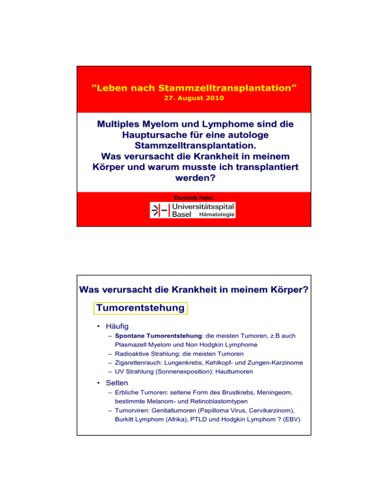
”Leben nach Stammzelltransplantation” 27. August 2010 Multiples Myelom und Lymphome sind die Hauptursache für eine autologe Stammzelltransplantation. Was verursacht die Krankheit in meinem Körper und warum musste ich transplantiert werden? Dominik Heim Hämatologie Was verursacht die Krankheit in meinem Körper? Tumorentstehung • Häufig – Spontane Tumorentstehung: die meisten Tumoren, z.B auch Plasmazell Myelom und Non Hodgkin Lymphome – Radioaktive Strahlung: die meisten Tumoren – Zigarettenrauch: Lungenkrebs, Kehlkopf- und Zungen-Karzinome – UV Strahlung (Sonnenexposition): Hauttumoren • Selten – Erbliche Tumoren: seltene Form des Brustkrebs, Meningeom, bestimmte Melanom- und Retinoblastomtypen – Tumorviren: Genitaltumoren (Papilloma Virus, Cervikarzinom), Burkitt Lymphom (Afrika), PTLD und Hodgkin Lymphom ? (EBV) Tumorentstehung Onkogene und Tumor Suppressor Gene Verlust der Funktion eines Tumor Suppressor Gens oder verstärkte Aktivität eines Onkogens führt zum Verlust des kontorllierten Zellwachstums Bsp. Tumor Suppressor Gen p53 Auswirkungen der Aktivierung des Bcr-Abl Onkoproteins Anti-apoptosis Was verursacht die Krankheit in meinem Körper? Tumorentstehung Erworbene genetische Veränderungen in den Tumorzellen • t(4;14) – typischerweise Multiples Myelom • t(14;16) – typisch Multiples Myelom • t(8;14) – typischerweise bei Burkitt-Lymphom, diffus-großzelligem B-NHL, selten Multiples Myelom • t(11;14) – typisch für das Mantelzelllymphom, gelegentlich Multiples Myelom oder chronische lymphatische Leukämie • t(14;18) – typisch für das follikuläre Lymphom • Del 17p – bei CLL, Multiplem Myelom • Del 11q – bei CLL DIFFERENZIERUNG UND PROLIFERATION (EXPANSION) ERYTHROZYTEN THROMBOZYTEN SELBSTERNEUERUNG GRANULOZYTEN MONOZYTEN STAMMZELLE T-LYMPHOZYTEN B-LYMPHOZYTEN DIFFERENZIERUNG DER B-ZELLEN CLL IgM D-J IgM IgD IgM IgG IgG/A IgG/A VDJ Stamm- pro-B pre-B “unreife “reife” Lympho- Memory Plasma- Plasmazelle ” B-Zelle blast B-Zelle blast zelle B-Zelle Ig Gen-Rearrangement Antigen unabhängig Ig class switch Antigen abhängig terminale Differenzierung Zweitkontakt MITOSE Autologe Stammzelltransplantation Lymphome • • Mature B cell neoplasms – Chronic lymphocytic leukemia – – – – B-cell prolymphocytic leukemia Lymphoplasmacytic lymphoma (such as Waldenström macroglobulinemia) Splenic marginal zone lymphoma Plasma cell neoplasms: • Plasma cell myeloma • • • Plasmacytoma Monoclonal immunoglobulin deposition diseases Heavy chain diseases – – Extranodal marginal zone B cell lymphoma, also called MALT lymphoma Nodal marginal zone B cell lymphoma (NMZL) – – – Follicular lymphoma Mantle cell lymphoma Diffuse large B cell lymphoma – – – – Mediastinal (thymic) large B cell lymphoma Intravascular large B cell lymphoma Primary effusion lymphoma Burkitt lymphoma/leukemia WHO-Klassifikation Mature T cell and natural killer (NK) cell neoplasms – – – – – – – – – – T cell prolymphocytic leukemia T cell large granular lymphocytic leukemia Aggressive NK cell leukemia Adult T cell leukemia/lymphoma Extranodal NK/T cell lymphoma, nasal type Enteropathy-type T cell lymphoma Hepatosplenic T cell lymphoma Blastic NK cell lymphoma Mycosis fungoides / Sezary syndrome Primary cutaneous CD30-positive T cell lymphoproliferative disorders – – – Angioimmunoblastic T cell lymphoma Peripheral T cell lymphoma, unspecified Anaplastic large cell lymphoma • • Primary cutaneous anaplastic large cell lymphoma Lymphomatoid papulosis Hodgkin lymphoma •Classical Hodgkin lymphomas: Nodular sclerosis Mixed cellularity Lymphocyte-rich Lymphocyte depleted or not depleted •Nodular lymphocyte-predominant Hodgkin lymphoma Therapie des Plasmazell Myeloms Historical perspective: 1962–2004 1962 Melphalan 1964 Cyclophosphamide 1967 Corticosteroids 1969 MP 1975 Durie-Salmon staging system 1982 Twin transplants for MM 1984 First allogeneic transplants; VAD 1985 IFN-α2 1996 Bisphosphonates 2000 Bortezomib; lenalidomide; other IMiDs®; Adriamycin® analogues; betathine; anti-angiogenics; arsenic 1976 First trials of complex chemotherapy combinations 1983 High-dose melphalan; serum β2-microglobulin for prognosis 1986 High-dose Dex; HDT with ACST 1996 first randomized study shows benefit of HDT-ASCT vs standard chemo 1999 Thalidomide; mini-allogeneic transplants 2001 New classification system 2004 International Staging System HDT with ASCT vs conventional chemotherapy Four published trials compared conventional chemotherapy (CC) with HDT in newly diagnosed Durie-Salmon stage II or III MM Age, years Study Attal et al.1 (IFM90) < 65 Fermand et al.2 (MAG91) 55–65 Blade et al.3 (PETHEMA) Median 56 Child et al.4 (MRC7) < 65 Median EFS, months Median OS, months Tx n CR, % CC 100 5* 18* 44* HDT 100 22* 28* 57* CC 96 – 19 47.6 HDT 94 – 25 47.8 CC 83 11* 33 66 HDT 81 30* 42 61 CC 200 8* 20* 42* HDT 201 44* 32* 54* * Significant p value. 1. Attal M, et al. N Engl J Med. 1996;335:91-97. 2. Fermand J, et al. J Clin Oncol. 2005;23:9227-33. 3. Blade J, et al. Blood. 2005;106:3755-59. 4. Child JA, et al. N Engl J Med. 2003;348:1875-83. Complete response rates with high-dose treatment How do the different stages of therapy contribute to remission? Induction High-dose therapy 100 90 CR rate (%) 80 70 60 50 40 30 20 10 0 Model of predicted response. ASCT Improved response rates Low treatment related mortality (1-2%) Prolonged EFS compared to standard therapy Prolonged OS (IFM and MRC trials) Novel agents as induction therapy for patients eligible for a transplant Conventional Thalidomide VAD Thal + Dex TAD CTD Dex Bortezomib VTD Vel + Dex PAD VCD Lenalidomide RVD RD Rd RAD Stem cell harvest High-dose melphalan Stem cell infusion C = cyclophosphamide; Rd = lenalidomide + low-dose dexamethasone; RD = lenalidomide + standard-dose dexamethasone. Complete response rates with high-dose treatment How do the different stages of therapy contribute to remission? Induction 100 90 CR rate (%) 80 70 60 Novel agents in induction 50 40 30 20 10 0 Model of predicted response. High-dose therapy Maintenance Plasmazell Myelom Klinik - Symptome Symptome bedingt durch Paraprotein Gewebeinfiltration Knochenkrankheit Symptomatisches PlasmazellMyelom definiert durch Paraprotein Klonale Plasmazellen CRAB C - hypercalcemia R - renal insufficiency + A - anemia B - bone lesions •hyperviscosity •amyloidosis •recurrent infections Sehstörungen Neurologische Sympt. Sympt. Blutungen Hyperviskosität Amyloidose Niereninsuffizienz Herzinsuffizienz andere Organe Niereninsuffizienz Cast Nephropathy CRAB Polyneuropathie Schmerzen Neuropathie 1. Paraprotein Gerinnungsstörung Solitäres Plasmozytom Blutungen Thrombosen Kompression Schmerzen Anämie CRAB Thrombopenie Verdrängung der Hämopoiese im Knochenmark Neutropenie 2. GewebeGewebe-Infiltration Hypogammaglobulinämie Knochenkrankheit Das MM ist der Tumor der am häufigsten das Skelett involviert – 90% der MM Patienten entwickeln Knochenläsionen – 60% der MM Patienten präsentieren sich mit Knochenschmerzen – 60% der MM Patienten entwickeln pathologische Frakturen MM Patienten verlieren rasch an Knochendichte – Patienten welche eine Steroid basierte Therapie erhalten verlieren in 1 Jahr bis zu 10% der Knochenmineralisation Osteolysen CRAB Schmerzen Pathologische Frakturen Querschnitt Symptomatik CRAB 3. Knochenkrankheit Polydipsie Polyurie Erbrechen Neurologische Symptome Koma Hyperkalzämie Niereninsuffizienz
