Aesculap Surgical Instruments TMT Instruments
Werbung
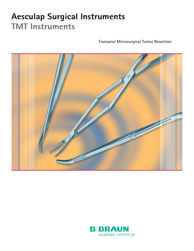
Aesculap Surgical Instruments TMT Instruments Transanal Microsurgical Tumor Resection TMT Instruments Instruments for Transanal Micorsurgical Tumor Resection (TMT) Instrumente für Transanale mikrochirurgische Tumorentfernung (TMT) Conventional techniques for the removal of rectal tumors (adenomas, polyps, small T1 carcinomas) have a number of unpleasant trouble for the patient, among them the possibility that a laparotomy or colostomy may have to be performed. For this reason an oncologic justified local resection is especially desirable. Transanal microsurgical tumor resection is a minimally invasive alternative technique, particularly for tumors at depths between 0 and 10 cm. Transanal access eliminates the need for laparotomy, and in many cases a colostomy and rectum extirpation can be avoided as well. The options offered by this technique range from the removal of polyps to the excision of tumors with full local intestinal wall resection and suturing. The operation technique is less invasive as a laparotomy and easier to perform than the endoscopic tumor resection (TEM). Specialized and easy to handle instruments have been developed for transanal tumor resection. A retractor with a cold light carrier provides additional illumination of the operative field, while special angled micro-instruments facilitate an unobstructed view of the surgical field. Thanks to a special transmission a greater depth (up to 14 cm) can be reached, than it would be possible with conventional instruments. Developed in cooperation with Professor Gottfried Müller, M.D. Caritas Krankenhaus Bad Mergentheim Uhlandstrasse 7 97980 Bad Mergentheim Die Entfernung von Tumoren (Adenome, Polypen, kleine T1 Karzinome) im Bereich des Rektums bringt bei der Verwendung konventioneller Operationstechniken für den Patienten vielfach Unannehmlichkeiten. Die Notwendigkeit einer Laparatomie und das Risiko eines künstlichen Darmausgangs sind hier zu nennen. Aus diesem Grund ist die onkologisch gerechtfertigte lokale Entfernung besonders erstrebenswert. Speziell für Tumore im Bereich von 0-10 cm Tiefe steht mit der Technik der transanalen mikrochirurgischen Entfernung von Tumoren eine patientenschonende Alternative zur Verfügung. Durch den transanalen Zugang entfällt die Laparatomie, ein künstlicher Darmausgang und Rektumextirpation kann in vielen Fällen vermieden werden. Die Möglichkeiten der Anwendungen reichen von der Entfernung von Polypen bis hin zur großflächigen Tumorentfernung mit Vollwandexzision und Naht. Hierbei ist die Operationstechnik weniger aufwendig als bei einer Laparatomie und einfacher als die endoskopische Tumorentfernung (TEM). Für die transanale mikrochirurgische Tumorentfernung wurde ein spezielles Instrumentarium entwickelt, das eine einfache Handhabung aufweist. Ein Sperrer mit Kaltlichtleiter bringt das notwendige Licht ins Operationsfeld. Speziell gewinkelte Mikroinstrumente ermöglichen eine ungehinderte Sicht auf den Operationsbereich. Durch eine spezielle Übersetzung wird eine größere Tiefe (bis maximal 14 cm) erreicht, als es mit konventionellen Instrumenten möglich wäre. Entwickelt in Zusammenarbeit mit: Prof. Dr. med. Gottfried Müller Caritas Krankenhaus Bad Mergentheim Uhlandstrasse 7 97980 Bad Mergentheim 1. Injecting saline solution with adrenaline (makes tumor larger and better to dissect, adrenaline will reduce bleeding) 1. Unterspritzen des Tumors mit NaCl und Adreanlin (der Tumor tritt besser hervor, das Adrenalin reduziert die Blutung) OP801 Cold light carrier, for additional, intensive illumination of the oprerating area Kaltlichtstab, zur zusätzlichen, intensiven Ausleuchtung des OP-Feldes OP906 OP913 OP914 Ø 4,8 mm 180 cm Ø 4,8 mm 250 cm Ø 4,8 mm 350 cm Fibre optic light cable, steamsterilizable up to 2 bar (134°C) Fiberglas-Lichtkabel, dampfsterilisierbar bis 2 bar (134°C) OP915J 100-110 Volt Japanese and South America version Ausführung für Japan und Südamerika OP915 220 Volt Light source, 150 watts, 5 degrees of brightness, possibility to switch-over to spare bulb Lichtquelle, 150 Watt, 5 Helligkeitsstufen, Umschaltmöglichkeit auf Reservelampe 2. Holding sutures are applied to the edges of the tumor. Thanks to the holding sutures the tumor can be manipulated and pulled down. 2. Haltefäden werden an den Tumorrändern angebracht. Dadurch kann der Tumor herabgezogen und bewegt werden. 3. Resection of the tumor and the surrounding mucous membrane. 3. Resektion des Tumors und der Darmschleimhaut. Ordering Data Bestelldaten BV589R PR = Pair/Paar BV591R BV581R OP801 BV582R BV591R OP906 - OP914 Center blade without connection for cold light carrier Center blade with connection for cold light carrier OP801 Mittelblatt ohne Aufnahme für Kaltlichtstab Mittelblatt mit Aufnahme für Kaltlichtstab OP801 BV581R BV583R BV584R BV586R BV589R Rectal spreader only Analsperrer allein 22 x 70 mm 22 x 93 mm 22 x 90 mm 23 x 150 mm Rectal blades Rektumblätter Sphincter blades Sphincterblätter for anastomosis für Anastomosen PR = Pair/Paar PR = Pair/Paar PR = Pair/Paar PARKS 4. Closure of the mucous membrane with sutures. A continous or single stitch suture can be applied. 4. Verschluss des Defektes der Darmschleimhaut mit Einzelknopf oder fortlaufender Naht. PR = Pair/Paar METZENBAUM EA921R Dissecting scissors Präparierschere EA922R Grasping Forceps Faßzange EA923R Needleholder Nadelhalter Aesculap AG & Co. KG Am Aesculap-Platz 78532 Tuttlingen Germany All rights reserved. Technical alterations are possible. This leaflet may be used for no other purposes than offering, buying and selling of our products. No part may be copied or reproduced in any form. In the case of misuse we retain the rights to recall our catalogues and pricelists and to take legal actions. Brochure No. C79411 Phone Fax +49 7461 95-0 +49 7461 95-2600 www.aesculap.de 0807/1.5/4
